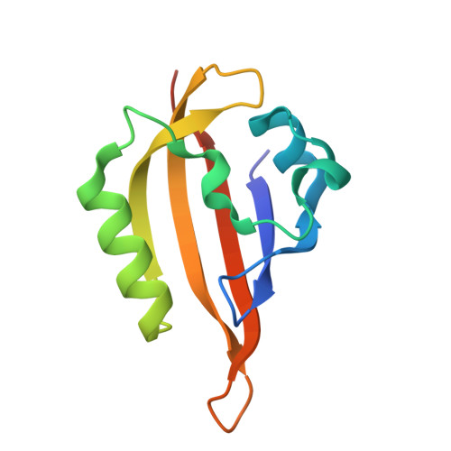
44OO | pdb_000044oo

Structure of Chloroflexus aggregans Cagg_3753 LOV domain C85A variant (CagFbFP) with 6,7-Dimethyl-8-ribityllumazine


Experimental Data Snapshot

  • Method: X-RAY DIFFRACTION
  • Resolution: 1.27 Å
  • R-Value Free: 
    0.203 (Depositor), 0.206 (DCC) 
  • R-Value Work: 
    0.174 (Depositor), 0.183 (DCC) 

Starting Model: experimental
View more details

wwPDB Validation 3D Report Full Report

Validation slider image for 44OO

Ligand Structure Quality Assessment 


This is version 1.1 of the entry. See complete history. 

Literature

Characterization of LOV domain-ribityllumazine complex with blue-shifted fluorescence.

Kuznetsova, E., Afanasev, A., Nikolaev, A., Natarov, I., Semenov, O., Mikhailov, A.E., Borshchevskiy, V., Yang, Y., Belozerova, O.A., Dubinnyi, M.A., Shlykov, M.A., Maksimov, E.G., Remeeva, A., Gushchin, I.

(2026) Biochem Biophys Res Commun 836: 154582-154582

  • DOI: https://doi.org/10.1016/j.bbrc.2026.154582
  • Primary Citation Related Structures: 
    44OO

  • PubMed Abstract: 

    Flavin-based fluorescent proteins (FbFPs) are small tags derived from natural LOV domains that can be used for anaerobic imaging. However, their applications are limited due to absence of strongly color-shifted variants. Here, we explore the possibility of reconstituting an FbFP with 6,7-dimethyl-8-ribityllumazine (LUM), an endogenous biosynthetic precursor of riboflavin (RF). We show that CagFbFP binds LUM with low-micromolar affinity (Kd = 9.5 ± 1.1 μM), forming a fluorescent complex with the excitation maximum at 419 nm and emission maximum at 462 nm. Under blue-light irradiation, CagFbFP-bound LUM is converted irreversibly to 6,7-dimethyllumazine, whereas the more traditional chromophore RF undergoes reversible reduction to hydroquinone form. Crystal structure of the CagFbFP-LUM complex determined at the anisotropic resolution reaching 1.27 Å confirms that the chromophore occupies the canonical flavin-binding pocket. The obtained results pave the way towards engineering of LUM-based FbFP variants with blue-shifted fluorescence emission.


  • Organizational Affiliation: 
    • Moscow Center for Advanced Studies, Moscow, 123592, Russia.

Macromolecule Content 

  • Total Structure Weight: 26.18 kDa 
  • Atom Count: 2,045 
  • Modeled Residue Count: 212 
  • Deposited Residue Count: 226 
  • Unique protein chains: 1

Macromolecules

Find similar proteins by:|  3D Structure
Entity ID: 1
MoleculeChains  Sequence LengthOrganismDetailsImage
Circadian input-output histidine kinase CikA
A, B
113Chloroflexus aggregans DSM 9485Mutation(s): 1 
Gene Names: Cagg_3753
EC: 2.7.13.3
UniProt
Find proteins for B8GAY9 (Chloroflexus aggregans (strain MD-66 / DSM 9485))
Explore B8GAY9 
Go to UniProtKB:  B8GAY9
Entity Groups
Sequence Clusters30% Identity50% Identity70% Identity90% Identity95% Identity100% Identity
UniProt GroupB8GAY9
Sequence Annotations
Expand
Reference Sequence

Small Molecules

Ligands 4 Unique
IDChains Name / Formula / InChI Key2D Diagram3D Interactions
DLZ
(Subject of Investigation/LOI)

Query on DLZ


Download:Ideal Coordinates CCD File
D [auth A],
H [auth B]
1-deoxy-1-(6,7-dimethyl-2,4-dioxo-3,4-dihydropteridin-8(2H)-yl)-D-ribitol
C13 H18 N4 O6
SXDXRJZUAJBNFL-XKSSXDPKSA-N
SO4

Query on SO4


Download:Ideal Coordinates CCD File
F [auth A],
G [auth A],
I [auth B],
J [auth B]
SULFATE ION
O4 S
QAOWNCQODCNURD-UHFFFAOYSA-L
GOL

Query on GOL


Download:Ideal Coordinates CCD File
C [auth A],
K [auth B],
L [auth B]
GLYCEROL
C3 H8 O3
PEDCQBHIVMGVHV-UHFFFAOYSA-N
EDO

Query on EDO


Download:Ideal Coordinates CCD File
E [auth A]1,2-ETHANEDIOL
C2 H6 O2
LYCAIKOWRPUZTN-UHFFFAOYSA-N

Experimental Data & Validation

Experimental Data

  • Method: X-RAY DIFFRACTION
  • Resolution: 1.27 Å
  • R-Value Free:  0.203 (Depositor), 0.206 (DCC) 
  • R-Value Work:  0.174 (Depositor), 0.183 (DCC) 
Space Group: P 21 21 2
Unit Cell:
Length ( Å )Angle ( ˚ )
a = 54.146α = 90
b = 110.352β = 90
c = 38.906γ = 90
Software Package:
Software NamePurpose
REFMACrefinement
XDSdata reduction
Aimlessdata scaling
MOLREPphasing

Structure Validation

View Full Validation Report


Ligand Structure Quality Assessment 


Entry History 

& Funding Information

Deposition Data


Funding OrganizationLocationGrant Number
Not funded--

Revision History  (Full details and data files)

  • Version 1.0: 2026-09-16
    Type: Initial release
  • Version 1.1: 2026-10-21
    Changes: Database references