3ZD1

STRUCTURE OF THE TWO C-TERMINAL DOMAINS OF COMPLEMENT FACTOR H RELATED PROTEIN 2


Experimental Data Snapshot

  • Method: X-RAY DIFFRACTION
  • Resolution: 2.00 Å
  • R-Value Free: 0.240 
  • R-Value Work: 0.211 
  • R-Value Observed: 0.212 

wwPDB Validation   3D Report Full Report


This is version 1.3 of the entry. See complete history


Literature

Dimerization of Complement Factor H-Related Proteins Modulates Complement Activation in Vivo.

Goicoechea De Jorge, E.Caesar, J.J.E.Malik, T.H.Patel, M.Colledge, M.Johnson, S.Hakobyan, S.Morgan, B.P.Harris, C.L.Pickering, M.C.Lea, S.M.

(2013) Proc Natl Acad Sci U S A 110: 4685

  • DOI: https://doi.org/10.1073/pnas.1219260110
  • Primary Citation of Related Structures:  
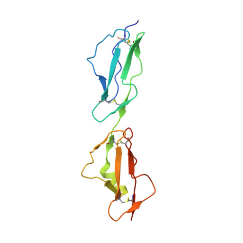
    3ZD1, 3ZD2

  • PubMed Abstract: 

    The complement system is a key component regulation influences susceptibility to age-related macular degeneration, meningitis, and kidney disease. Variation includes genomic rearrangements within the complement factor H-related (CFHR) locus. Elucidating the mechanism underlying these associations has been hindered by the lack of understanding of the biological role of CFHR proteins. Here we present unique structural data demonstrating that three of the CFHR proteins contain a shared dimerization motif and that this hitherto unrecognized structural property enables formation of both homodimers and heterodimers. Dimerization confers avidity for tissue-bound complement fragments and enables these proteins to efficiently compete with the physiological complement inhibitor, complement factor H (CFH), for ligand binding. Our data demonstrate that these CFHR proteins function as competitive antagonists of CFH to modulate complement activation in vivo and explain why variation in the CFHRs predisposes to disease.


  • Organizational Affiliation

    Centre for Complement and Inflammation Research, Department of Medicine, Imperial College, London W12 0NN, United Kingdom.


Macromolecules
Find similar proteins by:  (by identity cutoff)  |  3D Structure
Entity ID: 1
MoleculeChains Sequence LengthOrganismDetailsImage
COMPLEMENT FACTOR H-RELATED PROTEIN 2
A, B
126Homo sapiensMutation(s): 0 
UniProt & NIH Common Fund Data Resources
Find proteins for P36980 (Homo sapiens)
Explore P36980 
Go to UniProtKB:  P36980
PHAROS:  P36980
GTEx:  ENSG00000080910 
Entity Groups  
Sequence Clusters30% Identity50% Identity70% Identity90% Identity95% Identity100% Identity
UniProt GroupP36980
Sequence Annotations
Expand
  • Reference Sequence
Experimental Data & Validation

Experimental Data

  • Method: X-RAY DIFFRACTION
  • Resolution: 2.00 Å
  • R-Value Free: 0.240 
  • R-Value Work: 0.211 
  • R-Value Observed: 0.212 
  • Space Group: P 1 2 1
Unit Cell:
Length ( Å )Angle ( ˚ )
a = 52.95α = 90
b = 25.18β = 93.8
c = 95.68γ = 90
Software Package:
Software NamePurpose
BUSTERrefinement
XDSdata reduction
SCALAdata scaling
PHASERphasing

Structure Validation

View Full Validation Report


Entry History 

Deposition Data

Revision History  (Full details and data files)

  • Version 1.0: 2013-03-13
    Type: Initial release
  • Version 1.1: 2013-03-27
    Changes: Database references
  • Version 1.2: 2013-04-03
    Changes: Database references
  • Version 1.3: 2023-12-20
    Changes: Data collection, Database references, Derived calculations, Other, Refinement description