Structural insight into the tetramerization of an iterative ketoreductase siam through aromatic residues in the interfaces
Wang, H., Zhang, H., Zou, Y., Mi, Y., Lin, S., Xie, Z., Yan, Y., Zhang, H.(2014) PLoS One 9: e97996-e97996
- PubMed: 24901639
- DOI: https://doi.org/10.1371/journal.pone.0097996
- Primary Citation of Related Structures:
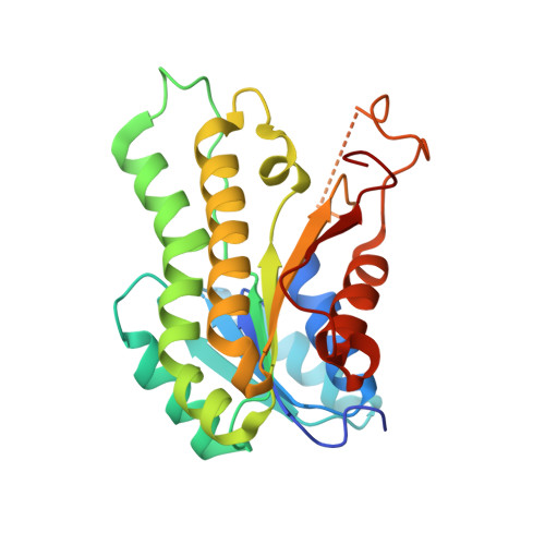
3WOH - PubMed Abstract:
In the biosynthesis of polyketides, ketoreductases (KRs) are an important group of enzymes that determine the chiralities of the carbon backbones. SiaM is a special member of this group that can recognize substrates with different lengths and can be used iteratively. Here we report the crystal structure of SiaM. Structural analysis indicates that the overall structure resembles those of other KRs. However, significant disparity can be found in the conserved LDD motif that is replaced with IRD motif in SiaM. The isoleucine and aspartic acid residues take similar orientations as leucine and aspartic acid in the conserved LDD motif, while the arginine residue points out towards the solvent. PISA analysis shows that SiaM forms a tetramer. Several aromatic residues are found in the interfaces, which have aromatic stacking interactions with the aromatic residues in the neighboring protomers. Mutagenesis studies performed on the aromatic residues show that these sites are important for maintaining the structural integrity of SiaM. However, the aromatic residues contribute differently to the enzymatic activity. In the N-terminal interface, the aromatic residues can be replaced with leucine without affecting the enzymatic activity while, in the other interface, such mutations abolish the enzymatic activity.
Organizational Affiliation:
Key Laboratory of Molecular Biophysics, Ministry of Education, Wuhan, Hubei, China; Department of Biotechnology, College of Life Science and Technology, Huazhong University of Science and Technology, Wuhan, Hubei, China.