3VWB

Crystal structure of VirB core domain (Se-Met derivative) complexed with the cis-acting site (5-BRU modifications) upstream icsb promoter


Experimental Data Snapshot

  • Method: X-RAY DIFFRACTION
  • Resolution: 2.42 Å
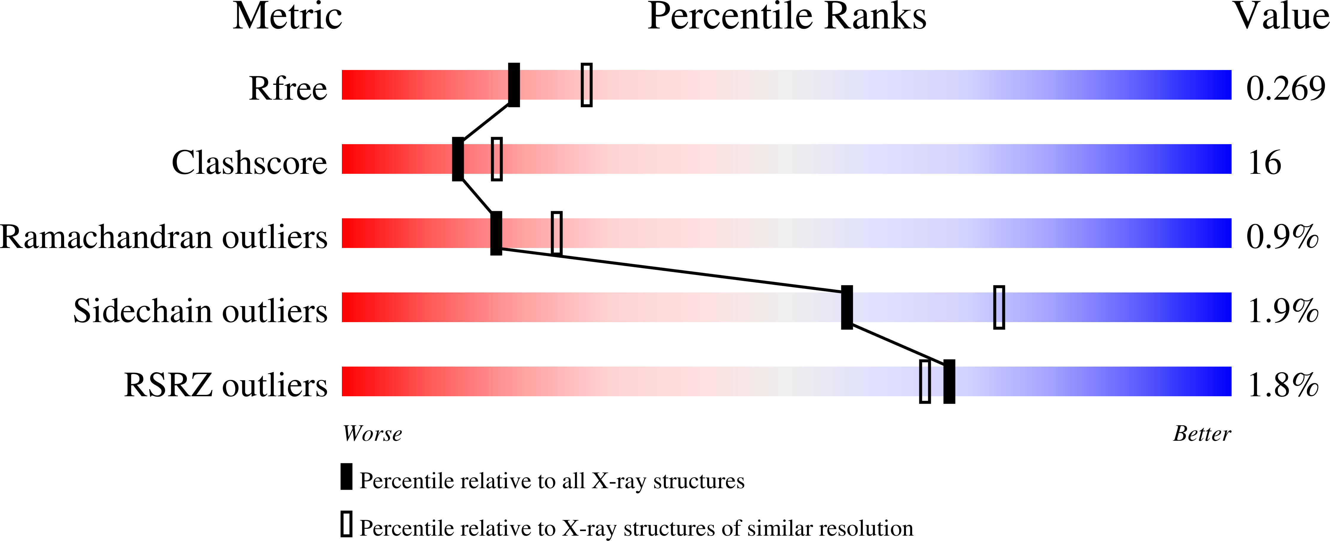
  • R-Value Free: 0.271 
  • R-Value Work: 0.235 
  • R-Value Observed: 0.237 

wwPDB Validation   3D Report Full Report


This is version 1.4 of the entry. See complete history


Literature

Structural insights into VirB-DNA complexes reveal mechanism of transcriptional activation of virulence genes

Gao, X.P.Zou, T.T.Mu, Z.X.Qin, B.Yang, J.Waltersperger, S.Wang, M.T.Cui, S.Jin, Q.

(2013) Nucleic Acids Res 41: 10529-10541

  • DOI: https://doi.org/10.1093/nar/gkt748
  • Primary Citation of Related Structures:  
    3VWB, 3W2A, 3W3C

  • PubMed Abstract: 

    VirB activates transcription of virulence genes in Shigella flexneri by alleviating heat-stable nucleoid-structuring protein-mediated promoter repression. VirB is unrelated to the conventional transcriptional regulators, but homologous to the plasmid partitioning proteins. We determined the crystal structures of VirB HTH domain bound by the cis-acting site containing the inverted repeat, revealing that the VirB-DNA complex is related to ParB-ParS-like complexes, presenting an example that a ParB-like protein acts exclusively in transcriptional regulation. The HTH domain of VirB docks DNA major groove and provides multiple contacts to backbone and bases, in which the only specific base readout is mediated by R167. VirB only recognizes one half site of the inverted repeats containing the most matches to the consensus for VirB binding. The binding of VirB induces DNA conformational changes and introduces a bend at an invariant A-tract segment in the cis-acting site, suggesting a role of DNA remodeling. VirB exhibits positive cooperativity in DNA binding that is contributed by the C-terminal domain facilitating VirB oligomerization. The isolated HTH domain only confers partial DNA specificity. Additional determinants for sequence specificity may reside in N- or C-terminal domains. Collectively, our findings support and extend a previously proposed model for relieving heat-stable nucleoid-structuring protein-mediated repression by VirB.


  • Organizational Affiliation

    MOH Key Laboratory of Systems Biology of Pathogens, Institute of Pathogen Biology, Chinese Academy of Medical Sciences & Peking Union Medical College, No.9 Dong Dan San Tiao, Beijing 100730, P.R. China and PX Beamlines Swiss Light Source at Paul Scherrer Institute, CH-5232 Villigen, Switzerland.


Macromolecules

Find similar proteins by:  (by identity cutoff)  |  3D Structure
Entity ID: 1
MoleculeChains Sequence LengthOrganismDetailsImage
Virulence regulon transcriptional activator virB143Shigella flexneri 2aMutation(s): 0 
Gene Names: virB
UniProt
Find proteins for P0A247 (Shigella flexneri)
Explore P0A247 
Go to UniProtKB:  P0A247
Entity Groups  
Sequence Clusters30% Identity50% Identity70% Identity90% Identity95% Identity100% Identity
UniProt GroupP0A247
Sequence Annotations
Expand
  • Reference Sequence
Find similar nucleic acids by:  (by identity cutoff)  |  3D Structure
Entity ID: 2
MoleculeChains LengthOrganismImage
DNA (26-MER)B [auth C]26N/A
Sequence Annotations
Expand
  • Reference Sequence
Find similar nucleic acids by:  (by identity cutoff)  |  3D Structure
Entity ID: 3
MoleculeChains LengthOrganismImage
DNA (26-MER)C [auth B]26N/A
Sequence Annotations
Expand
  • Reference Sequence
Experimental Data & Validation

Experimental Data

  • Method: X-RAY DIFFRACTION
  • Resolution: 2.42 Å
  • R-Value Free: 0.271 
  • R-Value Work: 0.235 
  • R-Value Observed: 0.237 
  • Space Group: P 21 21 2
Unit Cell:
Length ( Å )Angle ( ˚ )
a = 57.321α = 90
b = 163.051β = 90
c = 39.569γ = 90
Software Package:
Software NamePurpose
RemDAqdata collection
SHELXDphasing
SHELXEmodel building
PHENIXrefinement
XDSdata reduction
XDSdata scaling

Structure Validation

View Full Validation Report


Entry History 

Deposition Data

Revision History  (Full details and data files)

  • Version 1.0: 2013-08-21
    Type: Initial release
  • Version 1.1: 2013-09-04
    Changes: Database references, Structure summary
  • Version 1.2: 2013-12-11
    Changes: Database references
  • Version 1.3: 2013-12-25
    Changes: Database references
  • Version 1.4: 2024-03-20
    Changes: Data collection, Database references, Derived calculations