


MarR structures
Lou, H., Zhu, R., Hao, Z.To be published.
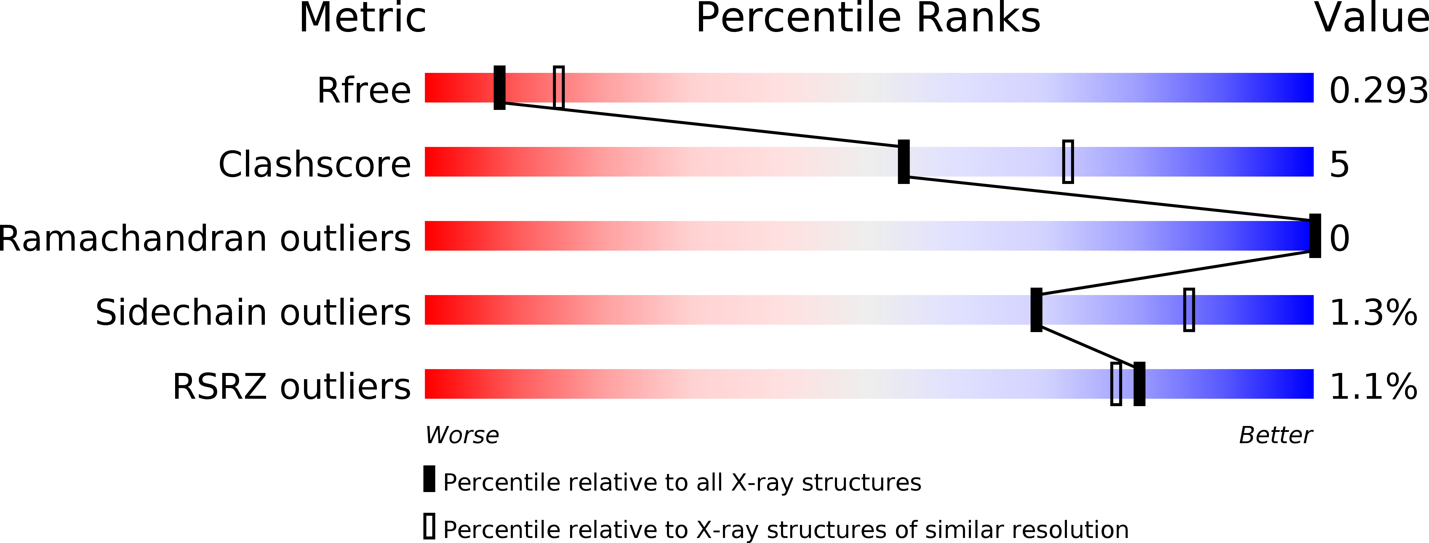
Experimental Data Snapshot
wwPDB Validation 3D Report Full Report
Entity ID: 1 | |||||
|---|---|---|---|---|---|
| Molecule | Chains | Sequence Length | Organism | Details | Image |
| Multiple antibiotic resistance protein marR | 144 | Escherichia coli K-12 | Mutation(s): 0 Gene Names: b1530, cfxB, inaR, JW5248, marR, soxQ | 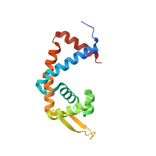 | |
UniProt | |||||
Find proteins for P27245 (Escherichia coli (strain K12)) Explore P27245 Go to UniProtKB: P27245 | |||||
Entity Groups | |||||
| Sequence Clusters | 30% Identity50% Identity70% Identity90% Identity95% Identity100% Identity | ||||
| UniProt Group | P27245 | ||||
Sequence AnnotationsExpand | |||||
| |||||
| Length ( Å ) | Angle ( ˚ ) |
|---|---|
| a = 61.96 | α = 90 |
| b = 61.96 | β = 90 |
| c = 131.74 | γ = 90 |
| Software Name | Purpose |
|---|---|
| MOSFLM | data reduction |
| SCALA | data scaling |
| PHASER | phasing |
| REFMAC | refinement |
| PDB_EXTRACT | data extraction |
RCSB PDB (citation) is hosted by
RCSB PDB is a member of the

