3VO0

Crystal structure of beta-glucuronidase from Acidobacterium capsulatum covalent-bonded with 2-deoxy-2-fluoro-D-glucuronic acid


Experimental Data Snapshot

  • Method: X-RAY DIFFRACTION
  • Resolution: 1.90 Å
  • R-Value Free: 0.240 
  • R-Value Work: 0.206 
  • R-Value Observed: 0.208 

wwPDB Validation   3D Report Full Report


Ligand Structure Quality Assessment 


This is version 2.1 of the entry. See complete history


Literature

Structural and biochemical characterization of glycoside hydrolase family 79 beta-glucuronidase from Acidobacterium capsulatum

Michikawa, M.Ichinose, H.Momma, M.Biely, P.Jongkees, S.Yoshida, M.Kotake, T.Tsumuraya, Y.Withers, S.G.Fujimoto, Z.Kaneko, S.

(2012) J Biol Chem 287: 14069-14077

  • DOI: https://doi.org/10.1074/jbc.M112.346288
  • Primary Citation of Related Structures:  
    3VNY, 3VNZ, 3VO0

  • PubMed Abstract: 

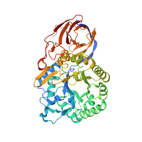
    We present the first structure of a glycoside hydrolase family 79 β-glucuronidase from Acidobacterium capsulatum, both as a product complex with β-D-glucuronic acid (GlcA) and as its trapped covalent 2-fluoroglucuronyl intermediate. This enzyme consists of a catalytic (β/α)(8)-barrel domain and a β-domain with irregular Greek key motifs that is of unknown function. The enzyme showed β-glucuronidase activity and trace levels of β-glucosidase and β-xylosidase activities. In conjunction with mutagenesis studies, these structures identify the catalytic residues as Glu(173) (acid base) and Glu(287) (nucleophile), consistent with the retaining mechanism demonstrated by (1)H NMR analysis. Glu(45), Tyr(243), Tyr(292)-Gly(294), and Tyr(334) form the catalytic pocket and provide substrate discrimination. Consistent with this, the Y292A mutation, which affects the interaction between the main chains of Gln(293) and Gly(294) and the GlcA carboxyl group, resulted in significant loss of β-glucuronidase activity while retaining the side activities at wild-type levels. Likewise, although the β-glucuronidase activity of the Y334F mutant is ~200-fold lower (k(cat)/K(m)) than that of the wild-type enzyme, the β-glucosidase activity is actually 3 times higher and the β-xylosidase activity is only 2.5-fold lower than the equivalent parameters for wild type, consistent with a role for Tyr(334) in recognition of the C6 position of GlcA. The involvement of Glu(45) in discriminating against binding of the O-methyl group at the C4 position of GlcA is revealed in the fact that the E45D mutant hydrolyzes PNP-β-GlcA approximately 300-fold slower (k(cat)/K(m)) than does the wild-type enzyme, whereas 4-O-methyl-GlcA-containing oligosaccharides are hydrolyzed only 7-fold slower.


  • Organizational Affiliation

    Food Biotechnology Division, National Agriculture and Food Research Organization Food Research Institute, 2-1-12 Kannondai, Tsukuba, Ibaraki 305-8642, Japan.


Macromolecules
Find similar proteins by:  (by identity cutoff)  |  3D Structure
Entity ID: 1
MoleculeChains Sequence LengthOrganismDetailsImage
beta-Glucuronidase488Acidobacterium capsulatum ATCC 51196Mutation(s): 0 
Gene Names: ACP_2665
EC: 3.2.1.31
UniProt
Find proteins for C1F2K5 (Acidobacterium capsulatum (strain ATCC 51196 / DSM 11244 / BCRC 80197 / JCM 7670 / NBRC 15755 / NCIMB 13165 / 161))
Explore C1F2K5 
Go to UniProtKB:  C1F2K5
Entity Groups  
Sequence Clusters30% Identity50% Identity70% Identity90% Identity95% Identity100% Identity
UniProt GroupC1F2K5
Sequence Annotations
Expand
  • Reference Sequence
Small Molecules
Ligands 4 Unique
IDChains Name / Formula / InChI Key2D Diagram3D Interactions
GUF
Query on GUF

Download Ideal Coordinates CCD File 
C [auth A],
E [auth A]
2-deoxy-2-fluoro-beta-D-glucopyranuronic acid
C6 H9 F O6
LDVYRSUUUDDWHG-LVBGKUAWSA-N
GUZ
Query on GUZ

Download Ideal Coordinates CCD File 
B [auth A]2-deoxy-2-fluoro-alpha-D-glucopyranuronic acid
C6 H9 F O6
LDVYRSUUUDDWHG-CLQWQSTFSA-N
DNF
Query on DNF

Download Ideal Coordinates CCD File 
D [auth A],
F [auth A],
G [auth A]
2,4-DINITROPHENOL
C6 H4 N2 O5
UFBJCMHMOXMLKC-UHFFFAOYSA-N
PO4
Query on PO4

Download Ideal Coordinates CCD File 
H [auth A],
I [auth A]
PHOSPHATE ION
O4 P
NBIIXXVUZAFLBC-UHFFFAOYSA-K
Experimental Data & Validation

Experimental Data

  • Method: X-RAY DIFFRACTION
  • Resolution: 1.90 Å
  • R-Value Free: 0.240 
  • R-Value Work: 0.206 
  • R-Value Observed: 0.208 
  • Space Group: I 41 2 2
Unit Cell:
Length ( Å )Angle ( ˚ )
a = 101.489α = 90
b = 101.489β = 90
c = 218.238γ = 90
Software Package:
Software NamePurpose
ADSCdata collection
REFMACrefinement
HKL-2000data reduction
HKL-2000data scaling
REFMACphasing

Structure Validation

View Full Validation Report


Ligand Structure Quality Assessment 


Entry History 

Deposition Data

Revision History  (Full details and data files)

  • Version 1.0: 2012-02-22
    Type: Initial release
  • Version 1.1: 2015-08-19
    Changes: Database references
  • Version 2.0: 2020-07-29
    Type: Remediation
    Reason: Carbohydrate remediation
    Changes: Atomic model, Data collection, Database references, Derived calculations
  • Version 2.1: 2023-11-08
    Changes: Data collection, Database references, Refinement description, Structure summary