3USB

Crystal Structure of Bacillus anthracis Inosine Monophosphate Dehydrogenase in the complex with IMP


Experimental Data Snapshot

  • Method: X-RAY DIFFRACTION
  • Resolution: 2.38 Å
  • R-Value Free: 0.201 
  • R-Value Work: 0.170 
  • R-Value Observed: 0.172 

wwPDB Validation   3D Report Full Report


Ligand Structure Quality Assessment 


This is version 1.5 of the entry. See complete history


Literature

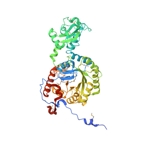
Bacillus anthracis inosine 5'-monophosphate dehydrogenase in action: the first bacterial series of structures of phosphate ion-, substrate-, and product-bound complexes.

Makowska-Grzyska, M.Kim, Y.Wu, R.Wilton, R.Gollapalli, D.R.Wang, X.K.Zhang, R.Jedrzejczak, R.Mack, J.C.Maltseva, N.Mulligan, R.Binkowski, T.A.Gornicki, P.Kuhn, M.L.Anderson, W.F.Hedstrom, L.Joachimiak, A.

(2012) Biochemistry 51: 6148-6163

  • DOI: https://doi.org/10.1021/bi300511w
  • Primary Citation of Related Structures:  
    3TSB, 3TSD, 3USB

  • PubMed Abstract: 

    Inosine 5'-monophosphate dehydrogenase (IMPDH) catalyzes the first unique step of the GMP branch of the purine nucleotide biosynthetic pathway. This enzyme is found in organisms of all three kingdoms. IMPDH inhibitors have broad clinical applications in cancer treatment, as antiviral drugs and as immunosuppressants, and have also displayed antibiotic activity. We have determined three crystal structures of Bacillus anthracis IMPDH, in a phosphate ion-bound (termed "apo") form and in complex with its substrate, inosine 5'-monophosphate (IMP), and product, xanthosine 5'-monophosphate (XMP). This is the first example of a bacterial IMPDH in more than one state from the same organism. Furthermore, for the first time for a prokaryotic enzyme, the entire active site flap, containing the conserved Arg-Tyr dyad, is clearly visible in the structure of the apoenzyme. Kinetic parameters for the enzymatic reaction were also determined, and the inhibitory effect of XMP and mycophenolic acid (MPA) has been studied. In addition, the inhibitory potential of two known Cryptosporidium parvum IMPDH inhibitors was examined for the B. anthracis enzyme and compared with those of three bacterial IMPDHs from Campylobacter jejuni, Clostridium perfringens, and Vibrio cholerae. The structures contribute to the characterization of the active site and design of inhibitors that specifically target B. anthracis and other microbial IMPDH enzymes.


  • Organizational Affiliation

    Center for Structural Genomics of Infectious Diseases, University of Chicago, 5735 South Ellis Avenue, Chicago, IL 60637, USA.


Macromolecules
Find similar proteins by:  (by identity cutoff)  |  3D Structure
Entity ID: 1
MoleculeChains Sequence LengthOrganismDetailsImage
Inosine-5'-monophosphate dehydrogenase
A, B
511Bacillus anthracis str. AmesMutation(s): 0 
Gene Names: guaBBA_0008BAS0011GBAA_0008
EC: 1.1.1.205
UniProt
Find proteins for A0A6L8P2U9 (Bacillus anthracis)
Explore A0A6L8P2U9 
Go to UniProtKB:  A0A6L8P2U9
Entity Groups  
Sequence Clusters30% Identity50% Identity70% Identity90% Identity95% Identity100% Identity
UniProt GroupA0A6L8P2U9
Sequence Annotations
Expand
  • Reference Sequence
Small Molecules
Ligands 4 Unique
IDChains Name / Formula / InChI Key2D Diagram3D Interactions
IMP
Query on IMP

Download Ideal Coordinates CCD File 
C [auth A],
G [auth B]
INOSINIC ACID
C10 H13 N4 O8 P
GRSZFWQUAKGDAV-KQYNXXCUSA-N
SO4
Query on SO4

Download Ideal Coordinates CCD File 
D [auth A],
J [auth B]
SULFATE ION
O4 S
QAOWNCQODCNURD-UHFFFAOYSA-L
GOL
Query on GOL

Download Ideal Coordinates CCD File 
E [auth A],
F [auth A],
H [auth B],
I [auth B]
GLYCEROL
C3 H8 O3
PEDCQBHIVMGVHV-UHFFFAOYSA-N
CL
Query on CL

Download Ideal Coordinates CCD File 
K [auth B]CHLORIDE ION
Cl
VEXZGXHMUGYJMC-UHFFFAOYSA-M
Modified Residues  1 Unique
IDChains TypeFormula2D DiagramParent
MSE
Query on MSE
A, B
L-PEPTIDE LINKINGC5 H11 N O2 SeMET
Experimental Data & Validation

Experimental Data

  • Method: X-RAY DIFFRACTION
  • Resolution: 2.38 Å
  • R-Value Free: 0.201 
  • R-Value Work: 0.170 
  • R-Value Observed: 0.172 
  • Space Group: I 4
  • Diffraction Data: https://doi.org/10.18430/M33USB
Unit Cell:
Length ( Å )Angle ( ˚ )
a = 122.519α = 90
b = 122.519β = 90
c = 140.9γ = 90
Software Package:
Software NamePurpose
PHENIXrefinement
HKL-3000data collection
HKL-3000phasing
SHELXSphasing
MLPHAREphasing
RESOLVEmodel building
SOLVEphasing
REFMACrefinement
SBC-Collectdata collection
HKL-3000data reduction
HKL-3000data scaling
RESOLVEphasing

Structure Validation

View Full Validation Report


Ligand Structure Quality Assessment 


Entry History 

Revision History  (Full details and data files)

  • Version 1.0: 2011-12-07
    Type: Initial release
  • Version 1.1: 2013-04-10
    Changes: Database references
  • Version 1.2: 2013-07-24
    Changes: Source and taxonomy
  • Version 1.3: 2018-01-24
    Changes: Structure summary
  • Version 1.4: 2019-07-17
    Changes: Data collection, Derived calculations, Refinement description
  • Version 1.5: 2019-08-14
    Changes: Data collection