3UE6

The dark structure of the blue-light photoreceptor Aureochrome1 LOV


Experimental Data Snapshot

  • Method: X-RAY DIFFRACTION
  • Resolution: 2.75 Å
  • R-Value Free: 0.256 
  • R-Value Work: 0.192 
  • R-Value Observed: 0.196 

wwPDB Validation   3D Report Full Report


Ligand Structure Quality Assessment 


This is version 1.4 of the entry. See complete history


Literature

Crystal structures of Aureochrome1 LOV suggest new design strategies for optogenetics.

Mitra, D.Yang, X.Moffat, K.

(2012) Structure 20: 698-706

  • DOI: https://doi.org/10.1016/j.str.2012.02.016
  • Primary Citation of Related Structures:  
    3UE6, 3ULF

  • PubMed Abstract: 

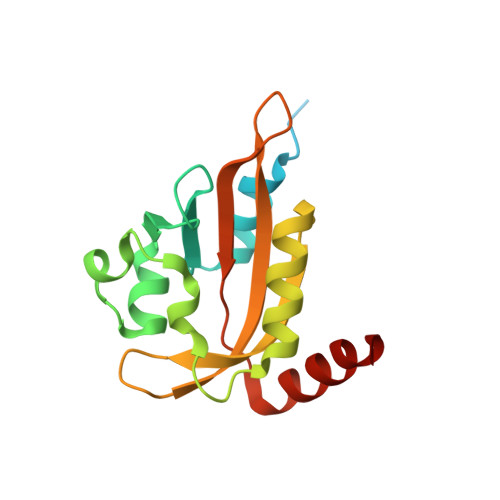
    Aureochrome1, a signaling photoreceptor from a eukaryotic photosynthetic stramenopile, confers blue-light-regulated DNA binding on the organism. Its topology, in which a C-terminal LOV sensor domain is linked to an N-terminal DNA-binding bZIP effector domain, contrasts with the reverse sensor-effector topology in most other known LOV-photoreceptors. How, then, is signal transmitted in Aureochrome1? The dark- and light-state crystal structures of Aureochrome1 LOV domain (AuLOV) show that its helical N- and C-terminal flanking regions are packed against the external surface of the core β sheet, opposite to the FMN chromophore on the internal surface. Light-induced conformational changes occur in the quaternary structure of the AuLOV dimer and in Phe298 of the Hβ strand in the core. The properties of AuLOV extend the applicability of LOV domains as versatile design modules that permit fusion to effector domains via either the N- or C-termini to confer blue-light sensitivity.


  • Organizational Affiliation

    Department of Biochemistry and Molecular Biology, University of Chicago, 929 E 57th Street, Chicago, IL 60637, USA.


Macromolecules
Find similar proteins by:  (by identity cutoff)  |  3D Structure
Entity ID: 1
MoleculeChains Sequence LengthOrganismDetailsImage
Aureochrome1
A, B, C, D, E
A, B, C, D, E, F
166Vaucheria frigidaMutation(s): 0 
Gene Names: AUREO1
UniProt
Find proteins for A8QW55 (Vaucheria frigida)
Explore A8QW55 
Go to UniProtKB:  A8QW55
Entity Groups  
Sequence Clusters30% Identity50% Identity70% Identity90% Identity95% Identity100% Identity
UniProt GroupA8QW55
Sequence Annotations
Expand
  • Reference Sequence
Experimental Data & Validation

Experimental Data

  • Method: X-RAY DIFFRACTION
  • Resolution: 2.75 Å
  • R-Value Free: 0.256 
  • R-Value Work: 0.192 
  • R-Value Observed: 0.196 
  • Space Group: P 43
Unit Cell:
Length ( Å )Angle ( ˚ )
a = 73.987α = 90
b = 73.987β = 90
c = 177.468γ = 90
Software Package:
Software NamePurpose
PHENIXrefinement
REFMACrefinement
HKL-2000data reduction
HKL-2000data scaling
PHASERphasing

Structure Validation

View Full Validation Report


Ligand Structure Quality Assessment 


Entry History 

Deposition Data

Revision History  (Full details and data files)

  • Version 1.0: 2012-04-11
    Type: Initial release
  • Version 1.1: 2013-02-20
    Changes: Database references
  • Version 1.2: 2017-11-08
    Changes: Refinement description
  • Version 1.3: 2019-07-17
    Changes: Data collection, Refinement description
  • Version 1.4: 2024-02-28
    Changes: Data collection, Database references, Derived calculations