3UBR

Laue structure of Shewanella oneidensis cytochrome-c Nitrite Reductase


Experimental Data Snapshot

  • Method: X-RAY DIFFRACTION
  • Resolution: 2.59 Å
  • R-Value Free: 0.285 
  • R-Value Work: 0.190 
  • R-Value Observed: 0.195 

wwPDB Validation   3D Report Full Report


Ligand Structure Quality Assessment 


This is version 1.4 of the entry. See complete history


Literature

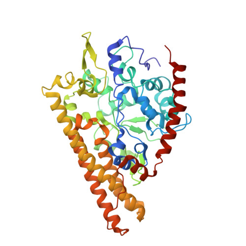
Laue crystal structure of Shewanella oneidensis cytochrome c nitrite reductase from a high-yield expression system.

Youngblut, M.Judd, E.T.Srajer, V.Sayyed, B.Goelzer, T.Elliott, S.J.Schmidt, M.Pacheco, A.A.

(2012) J Biol Inorg Chem 17: 647-662

  • DOI: https://doi.org/10.1007/s00775-012-0885-0
  • Primary Citation of Related Structures:  
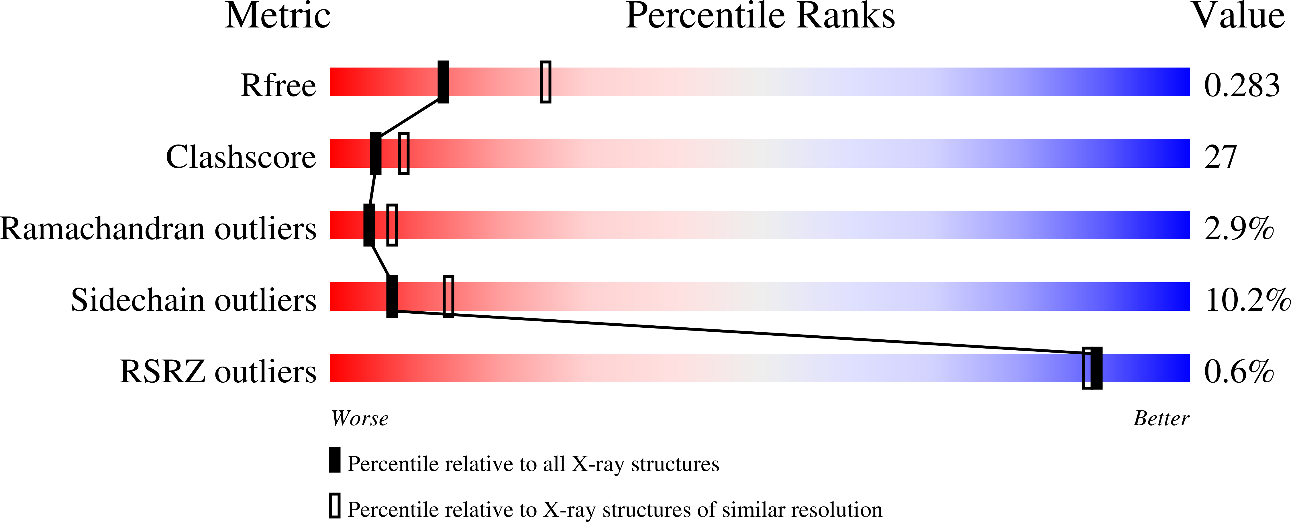
    3UBR

  • PubMed Abstract: 

    The high-yield expression and purification of Shewanella oneidensis cytochrome c nitrite reductase (ccNiR) and its characterization by a variety of methods, notably Laue crystallography, are reported. A key component of the expression system is an artificial ccNiR gene in which the N-terminal signal peptide from the highly expressed S. oneidensis protein "small tetraheme c" replaces the wild-type signal peptide. This gene, inserted into the plasmid pHSG298 and expressed in S. oneidensis TSP-1 strain, generated approximately 20 mg crude ccNiR per liter of culture, compared with 0.5-1 mg/L for untransformed cells. Purified ccNiR has nitrite and hydroxylamine reductase activities comparable to those previously reported for Escherichia coli ccNiR, and is stable for over 2 weeks in pH 7 solution at 4 °C. UV/vis spectropotentiometric titrations and protein film voltammetry identified five independent one-electron reduction processes. Global analysis of the spectropotentiometric data also allowed determination of the extinction coefficient spectra for the five reduced ccNiR species. The characteristics of the individual extinction coefficient spectra suggest that, within each reduced species, the electrons are distributed among the various hemes, rather than being localized on specific heme centers. The purified ccNiR yielded good-quality crystals, with which the 2.59-Å-resolution structure was solved at room temperature using the Laue diffraction method. The structure is similar to that of E. coli ccNiR, except in the region where the enzyme interacts with its physiological electron donor (CymA in the case of S. oneidensis ccNiR, NrfB in the case of the E. coli protein).


  • Organizational Affiliation

    Department of Chemistry and Biochemistry, University of Wisconsin-Milwaukee, 3210 N. Cramer St, Milwaukee, WI 53211, USA.


Macromolecules
Find similar proteins by:  (by identity cutoff)  |  3D Structure
Entity ID: 1
MoleculeChains Sequence LengthOrganismDetailsImage
Cytochrome c-552
A, B
439Shewanella oneidensisMutation(s): 0 
Gene Names: nrfAShewanella oneidensisSO_3980
EC: 1.7.2.2
UniProt
Find proteins for Q8EAC7 (Shewanella oneidensis (strain MR-1))
Explore Q8EAC7 
Go to UniProtKB:  Q8EAC7
Entity Groups  
Sequence Clusters30% Identity50% Identity70% Identity90% Identity95% Identity100% Identity
UniProt GroupQ8EAC7
Sequence Annotations
Expand
  • Reference Sequence
Small Molecules
Ligands 2 Unique
IDChains Name / Formula / InChI Key2D Diagram3D Interactions
HEC
Query on HEC

Download Ideal Coordinates CCD File 
C [auth A]
D [auth A]
E [auth A]
F [auth A]
G [auth A]
C [auth A],
D [auth A],
E [auth A],
F [auth A],
G [auth A],
I [auth B],
J [auth B],
K [auth B],
L [auth B],
M [auth B]
HEME C
C34 H34 Fe N4 O4
HXQIYSLZKNYNMH-LJNAALQVSA-N
CA
Query on CA

Download Ideal Coordinates CCD File 
H [auth A],
N [auth B]
CALCIUM ION
Ca
BHPQYMZQTOCNFJ-UHFFFAOYSA-N
Experimental Data & Validation

Experimental Data

  • Method: X-RAY DIFFRACTION
  • Resolution: 2.59 Å
  • R-Value Free: 0.285 
  • R-Value Work: 0.190 
  • R-Value Observed: 0.195 
  • Space Group: P 21 21 21
Unit Cell:
Length ( Å )Angle ( ˚ )
a = 51.5α = 90
b = 95.9β = 90
c = 223.8γ = 90
Software Package:
Software NamePurpose
LaueCollectdata collection
PHASERphasing
REFMACrefinement
Precognitiondata reduction
Epinormdata reduction

Structure Validation

View Full Validation Report


Ligand Structure Quality Assessment 


Entry History 

Deposition Data

Revision History  (Full details and data files)

  • Version 1.0: 2012-04-25
    Type: Initial release
  • Version 1.1: 2012-06-20
    Changes: Database references
  • Version 1.2: 2017-03-15
    Changes: Other
  • Version 1.3: 2019-09-04
    Changes: Data collection, Derived calculations
  • Version 1.4: 2023-09-13
    Changes: Data collection, Database references, Derived calculations, Refinement description