3T4A

Structure of a truncated form of Staphylococcal Complement Inhibitor B bound to human C3c at 3.4 Angstrom resolution


Experimental Data Snapshot

  • Method: X-RAY DIFFRACTION
  • Resolution: 3.40 Å
  • R-Value Free: 0.270 
  • R-Value Work: 0.229 
  • R-Value Observed: 0.231 

wwPDB Validation   3D Report Full Report


This is version 1.4 of the entry. See complete history


Literature

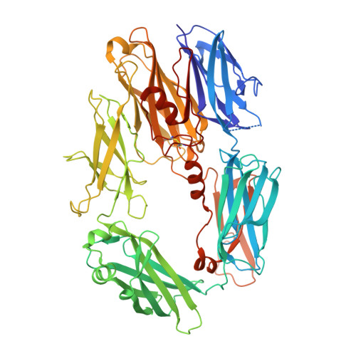
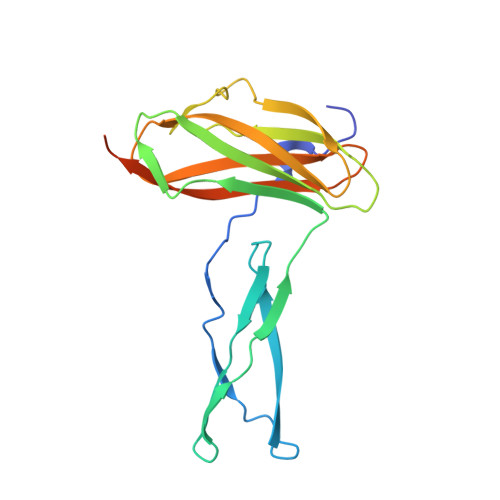
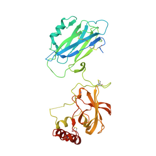
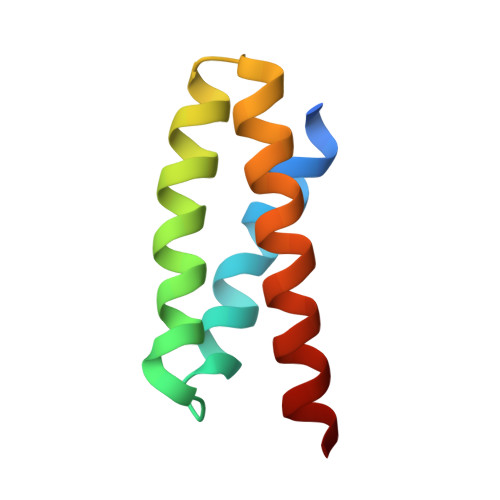
Diversity in the C3b Convertase Contact Residues and Tertiary Structures of the Staphylococcal Complement Inhibitor (SCIN) Protein Family.

Garcia, B.L.Summers, B.J.Lin, Z.Ramyar, K.X.Ricklin, D.Kamath, D.V.Fu, Z.Q.Lambris, J.D.Geisbrecht, B.V.

(2012) J Biol Chem 287: 628-640

  • DOI: https://doi.org/10.1074/jbc.M111.298984
  • Primary Citation of Related Structures:  
    3T46, 3T47, 3T48, 3T49, 3T4A

  • PubMed Abstract: 

    To survive in immune-competent hosts, the pathogen Staphylococcus aureus expresses and secretes a sophisticated array of proteins that inhibit the complement system. Among these are the staphylococcal complement inhibitors (SCIN), which are composed of three active proteins (SCIN-A, -B, and -C) and one purportedly inactive member (SCIN-D or ORF-D). Because previous work has focused almost exclusively on SCIN-A, we sought to provide initial structure/function information on additional SCIN proteins. To this end we determined crystal structures of an active, N-terminal truncation mutant of SCIN-B (denoted SCIN-B18-85) both free and bound to the C3c fragment of complement component C3 at 1.5 and 3.4 Å resolution, respectively. Comparison of the C3c/SCIN-B18-85 structure with that of C3c/SCIN-A revealed that both proteins target the same functional hotspot on the C3b/C3c surface yet harbor diversity in both the type of residues and interactions formed at their C3b/C3c interfaces. Most importantly, these structures allowed identification of Arg44 and Tyr51 as residues key for SCIN-B binding to C3b and subsequent inhibition of the AP C3 convertase. In addition, we also solved several crystal structures of SCIN-D to 1.3 Å limiting resolution. This revealed an unexpected structural deviation in the N-terminal α helix relative to SCIN-A and SCIN-B. Comparative analysis of both electrostatic potentials and surface complementarity suggest a physical explanation for the inability of SCIN-D to bind C3b/C3c. Together, these studies provide a more thorough understanding of immune evasion by S. aureus and enhance potential use of SCIN proteins as templates for design of complement targeted therapeutics.


  • Organizational Affiliation

    Division of Cell Biology and Biophysics, School of Biological Sciences, University of Missouri-Kansas City, Kansas City, Missouri 64110.


Macromolecules
Find similar proteins by:  (by identity cutoff)  |  3D Structure
Entity ID: 1
MoleculeChains Sequence LengthOrganismDetailsImage
Complement C3 beta chain
A, D
645Homo sapiensMutation(s): 0 
UniProt & NIH Common Fund Data Resources
Find proteins for P01024 (Homo sapiens)
Explore P01024 
Go to UniProtKB:  P01024
PHAROS:  P01024
GTEx:  ENSG00000125730 
Entity Groups  
Sequence Clusters30% Identity50% Identity70% Identity90% Identity95% Identity100% Identity
UniProt GroupP01024
Sequence Annotations
Expand
  • Reference Sequence
Find similar proteins by:  (by identity cutoff)  |  3D Structure
Entity ID: 2
MoleculeChains Sequence LengthOrganismDetailsImage
Complement C3c alpha' chain fragment 1
B, E
206Homo sapiensMutation(s): 0 
UniProt & NIH Common Fund Data Resources
Find proteins for P01024 (Homo sapiens)
Explore P01024 
Go to UniProtKB:  P01024
PHAROS:  P01024
GTEx:  ENSG00000125730 
Entity Groups  
Sequence Clusters30% Identity50% Identity70% Identity90% Identity95% Identity100% Identity
UniProt GroupP01024
Sequence Annotations
Expand
  • Reference Sequence
Find similar proteins by:  (by identity cutoff)  |  3D Structure
Entity ID: 3
MoleculeChains Sequence LengthOrganismDetailsImage
Complement C3c alpha' chain fragment 2
C, F
343Homo sapiensMutation(s): 0 
UniProt & NIH Common Fund Data Resources
Find proteins for P01024 (Homo sapiens)
Explore P01024 
Go to UniProtKB:  P01024
PHAROS:  P01024
GTEx:  ENSG00000125730 
Entity Groups  
Sequence Clusters30% Identity50% Identity70% Identity90% Identity95% Identity100% Identity
UniProt GroupP01024
Sequence Annotations
Expand
  • Reference Sequence
Find similar proteins by:  (by identity cutoff)  |  3D Structure
Entity ID: 4
MoleculeChains Sequence LengthOrganismDetailsImage
Fibrinogen-binding protein
G, H
73Staphylococcus aureus subsp. aureus Mu50Mutation(s): 0 
Gene Names: SAV1159
UniProt
Find proteins for A0A0H3JZ23 (Staphylococcus aureus (strain Mu50 / ATCC 700699))
Explore A0A0H3JZ23 
Go to UniProtKB:  A0A0H3JZ23
Entity Groups  
Sequence Clusters30% Identity50% Identity70% Identity90% Identity95% Identity100% Identity
UniProt GroupA0A0H3JZ23
Sequence Annotations
Expand
  • Reference Sequence
Experimental Data & Validation

Experimental Data

  • Method: X-RAY DIFFRACTION
  • Resolution: 3.40 Å
  • R-Value Free: 0.270 
  • R-Value Work: 0.229 
  • R-Value Observed: 0.231 
  • Space Group: P 21 21 21
Unit Cell:
Length ( Å )Angle ( ˚ )
a = 103.304α = 90
b = 165.436β = 90
c = 203.125γ = 90
Software Package:
Software NamePurpose
DENZOdata reduction
SCALEPACKdata scaling
PHASERphasing
PHENIXrefinement
PDB_EXTRACTdata extraction

Structure Validation

View Full Validation Report


Entry History 

Deposition Data

Revision History  (Full details and data files)

  • Version 1.0: 2011-11-23
    Type: Initial release
  • Version 1.1: 2012-01-18
    Changes: Database references
  • Version 1.2: 2013-05-22
    Changes: Refinement description
  • Version 1.3: 2017-11-08
    Changes: Refinement description
  • Version 1.4: 2023-09-13
    Changes: Data collection, Database references, Refinement description