3SPD

Crystal structure of aprataxin ortholog Hnt3 in complex with DNA


Experimental Data Snapshot

  • Method: X-RAY DIFFRACTION
  • Resolution: 1.91 Å
  • R-Value Free: 0.211 
  • R-Value Work: 0.175 
  • R-Value Observed: 0.176 

wwPDB Validation   3D Report Full Report


This is version 1.2 of the entry. See complete history


Literature

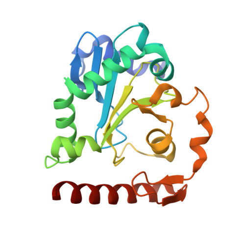
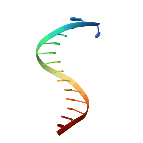
Crystal structures of aprataxin ortholog Hnt3 reveal the mechanism for reversal of 5'-adenylated DNA

Gong, Y.Zhu, D.Ding, J.Dou, C.Ren, X.Gu, L.Jiang, T.Wang, D.

(2011) Nat Struct Mol Biol 18: 1297-1299

  • DOI: https://doi.org/10.1038/nsmb.2145
  • Primary Citation of Related Structures:  
    3SP4, 3SPD, 3SPL

  • PubMed Abstract: 

    Aprataxin is a DNA deadenylase that resolves DNA 5'-AMP termini and reverses abortive DNA ligation. The crystal structures of Schizosaccharomyces pombe aprataxin Hnt3 in its apo form and in complex to dsDNA and dsDNA-AMP reveal how Hnt3 recognizes and processes 5'-adenylated DNA in a structure-specific manner. The bound DNA adopts a 5'-flap conformation that facilitates 5'-AMP access to the active site, where AMP cleavage occurs by a canonical catalytic mechanism.


  • Organizational Affiliation

    National Laboratory of Biomacromolecules, Institute of Biophysics, Chinese Academy of Sciences, Beijing, China.


Macromolecules

Find similar proteins by:  (by identity cutoff)  |  3D Structure
Entity ID: 1
MoleculeChains Sequence LengthOrganismDetailsImage
Aprataxin-like protein
A, B, C, D
204Schizosaccharomyces pombe 972h-Mutation(s): 1 
Gene Names: hnt3SPCC18.09c
UniProt
Find proteins for O74859 (Schizosaccharomyces pombe (strain 972 / ATCC 24843))
Explore O74859 
Go to UniProtKB:  O74859
Entity Groups  
Sequence Clusters30% Identity50% Identity70% Identity90% Identity95% Identity100% Identity
UniProt GroupO74859
Sequence Annotations
Expand
  • Reference Sequence

Find similar nucleic acids by:  Sequence   |   3D Structure  

Entity ID: 2
MoleculeChains LengthOrganismImage
DNA (5'-D(*TP*AP*TP*TP*CP*CP*GP*AP*TP*AP*GP*TP*GP*AP*C)-3')
E, G
15N/A
Sequence Annotations
Expand
  • Reference Sequence

Find similar nucleic acids by:  Sequence   |   3D Structure  

Entity ID: 3
MoleculeChains LengthOrganismImage
DNA (5'-D(*GP*TP*CP*AP*CP*TP*AP*TP*CP*GP*GP*AP*AP*TP*GP*AP*G)-3')
F, H
17N/A
Sequence Annotations
Expand
  • Reference Sequence
Small Molecules
Ligands 2 Unique
IDChains Name / Formula / InChI Key2D Diagram3D Interactions
SO4
Query on SO4

Download Ideal Coordinates CCD File 
AA [auth D]
BA [auth D]
J [auth A]
K [auth A]
L [auth A]
AA [auth D],
BA [auth D],
J [auth A],
K [auth A],
L [auth A],
M [auth A],
O [auth B],
P [auth B],
Q [auth B],
R [auth B],
T [auth C],
U [auth C],
V [auth C],
W [auth C],
Y [auth D],
Z [auth D]
SULFATE ION
O4 S
QAOWNCQODCNURD-UHFFFAOYSA-L
ZN
Query on ZN

Download Ideal Coordinates CCD File 
I [auth A],
N [auth B],
S [auth C],
X [auth D]
ZINC ION
Zn
PTFCDOFLOPIGGS-UHFFFAOYSA-N
Experimental Data & Validation

Experimental Data

  • Method: X-RAY DIFFRACTION
  • Resolution: 1.91 Å
  • R-Value Free: 0.211 
  • R-Value Work: 0.175 
  • R-Value Observed: 0.176 
  • Space Group: H 3 2
Unit Cell:
Length ( Å )Angle ( ˚ )
a = 220.409α = 90
b = 220.409β = 90
c = 135.031γ = 120
Software Package:
Software NamePurpose
HKL-2000data collection
PHASERphasing
PHENIXrefinement
HKL-2000data reduction
HKL-2000data scaling

Structure Validation

View Full Validation Report


Entry History 

Deposition Data

Revision History  (Full details and data files)

  • Version 1.0: 2011-10-12
    Type: Initial release
  • Version 1.1: 2013-07-03
    Changes: Database references
  • Version 1.2: 2023-11-01
    Changes: Data collection, Database references, Derived calculations, Refinement description