3RWL

Structure of P450pyr hydroxylase


Experimental Data Snapshot

  • Method: X-RAY DIFFRACTION
  • Resolution: 2.00 Å
  • R-Value Free: 0.196 
  • R-Value Work: 0.160 
  • R-Value Observed: 0.162 

wwPDB Validation   3D Report Full Report


Ligand Structure Quality Assessment 


This is version 1.2 of the entry. See complete history


Literature

Evolving P450pyr hydroxylase for highly enantioselective hydroxylation at non-activated carbon atom.

Pham, S.Q.Pompidor, G.Liu, J.Li, X.D.Li, Z.

(2012) Chem Commun (Camb) 48: 4618-4620

  • DOI: https://doi.org/10.1039/c2cc30779k
  • Primary Citation of Related Structures:  
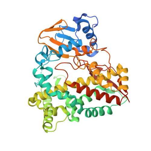
    3RWL

  • PubMed Abstract: 

    Directed evolution of a monooxygenase to achieve very high enantioselectivity for hydroxylation at non-activated carbon atoms is demonstrated for the first time, where a triple mutant of P450pyr hydroxylase is obtained via determination of enzyme structure, iterative saturation mutagenesis, and high-throughput screening with a MS-based ee assay to increase the product ee from 53% to 98% for the hydroxylation of N-benzyl pyrrolidine to (S)-N-benzyl 3-hydroxypyrrolidine.


  • Organizational Affiliation

    Department of Chemical and Biomolecular Engineering, National University of Singapore, 4 Engineering Drive 4, Singapore 117576.


Macromolecules
Find similar proteins by:  (by identity cutoff)  |  3D Structure
Entity ID: 1
MoleculeChains Sequence LengthOrganismDetailsImage
Cytochrome P450 alkane hydroxylase 1 CYP153A7426Sphingopyxis macrogoltabidaMutation(s): 0 
Gene Names: ahpG1Sphingomonas sp.
EC: 1.14.15.3
UniProt
Find proteins for Q5F4D9 (Sphingopyxis macrogoltabida)
Explore Q5F4D9 
Go to UniProtKB:  Q5F4D9
Entity Groups  
Sequence Clusters30% Identity50% Identity70% Identity90% Identity95% Identity100% Identity
UniProt GroupQ5F4D9
Sequence Annotations
Expand
  • Reference Sequence
Small Molecules
Ligands 1 Unique
IDChains Name / Formula / InChI Key2D Diagram3D Interactions
HEM
Query on HEM

Download Ideal Coordinates CCD File 
B [auth A]PROTOPORPHYRIN IX CONTAINING FE
C34 H32 Fe N4 O4
KABFMIBPWCXCRK-RGGAHWMASA-L
Experimental Data & Validation

Experimental Data

  • Method: X-RAY DIFFRACTION
  • Resolution: 2.00 Å
  • R-Value Free: 0.196 
  • R-Value Work: 0.160 
  • R-Value Observed: 0.162 
  • Space Group: P 31
Unit Cell:
Length ( Å )Angle ( ˚ )
a = 112.604α = 90
b = 112.604β = 90
c = 41.964γ = 120
Software Package:
Software NamePurpose
SCALAdata scaling
SHELXphasing
DMphasing
REFMACrefinement
PDB_EXTRACTdata extraction
XSCALEdata scaling
SHELXDphasing

Structure Validation

View Full Validation Report


Ligand Structure Quality Assessment 


Entry History 

Deposition Data

  • Released Date: 2012-04-18 
  • Deposition Author(s): Pompidor, G.

Revision History  (Full details and data files)

  • Version 1.0: 2012-04-18
    Type: Initial release
  • Version 1.1: 2012-04-25
    Changes: Database references
  • Version 1.2: 2024-02-28
    Changes: Data collection, Database references, Derived calculations