3QK2

Structure-Based Analysis of the Interaction between the Simian Virus 40 T-Antigen Origin Binding Domain and Single-Stranded DNA


Experimental Data Snapshot

  • Method: X-RAY DIFFRACTION
  • Resolution: 1.64 Å
  • R-Value Free: 0.187 
  • R-Value Work: 0.164 
  • R-Value Observed: 0.165 

wwPDB Validation   3D Report Full Report


This is version 1.2 of the entry. See complete history


Literature

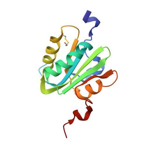
Structure-based analysis of the interaction between the simian virus 40 T-antigen origin binding domain and single-stranded DNA.

Meinke, G.Phelan, P.J.Fradet-Turcotte, A.Bohm, A.Archambault, J.Bullock, P.A.

(2011) J Virol 85: 818-827

  • DOI: https://doi.org/10.1128/JVI.01738-10
  • Primary Citation of Related Structures:  
    3QK2, 5D9I

  • PubMed Abstract: 

    The origin-binding domain (OBD) of simian virus 40 (SV40) large T-antigen (T-Ag) is essential for many of T-Ag's interactions with DNA. Nevertheless, many important issues related to DNA binding, for example, how single-stranded DNA (ssDNA) transits along the T-Ag OBD, have yet to be established. Therefore, X-ray crystallography was used to determine the costructure of the T-Ag OBD bound to DNA substrates such as the single-stranded region of a forked oligonucleotide. A second structure of the T-Ag OBD crystallized in the presence of poly(dT)(12) is also reported. To test the conclusions derived from these structures, residues identified as being involved in binding to ssDNA by crystallography or by an earlier nuclear magnetic resonance study were mutated, and their binding to DNA was characterized via fluorescence anisotropy. In addition, these mutations were introduced into full-length T-Ag, and these mutants were tested for their ability to support replication. When considered in terms of additional homology-based sequence alignments, our studies refine our understanding of how the T-Ag OBDs encoded by the polyomavirus family interact with ssDNA, a critical step during the initiation of DNA replication.


  • Organizational Affiliation

    Department of Biochemistry, Tufts University School of Medicine, 136 Harrison Avenue, Boston, MA 02111, USA.


Macromolecules
Find similar proteins by:  (by identity cutoff)  |  3D Structure
Entity ID: 1
MoleculeChains Sequence LengthOrganismDetailsImage
Large T antigen134Betapolyomavirus macacaeMutation(s): 0 
EC: 3.6.4
UniProt
Find proteins for P03070 (Simian virus 40)
Explore P03070 
Go to UniProtKB:  P03070
Entity Groups  
Sequence Clusters30% Identity50% Identity70% Identity90% Identity95% Identity100% Identity
UniProt GroupP03070
Sequence Annotations
Expand
  • Reference Sequence
Small Molecules
Ligands 1 Unique
IDChains Name / Formula / InChI Key2D Diagram3D Interactions
SCN
Query on SCN

Download Ideal Coordinates CCD File 
B [auth A]THIOCYANATE ION
C N S
ZMZDMBWJUHKJPS-UHFFFAOYSA-M
Modified Residues  1 Unique
IDChains TypeFormula2D DiagramParent
CME
Query on CME
A
L-PEPTIDE LINKINGC5 H11 N O3 S2CYS
Experimental Data & Validation

Experimental Data

  • Method: X-RAY DIFFRACTION
  • Resolution: 1.64 Å
  • R-Value Free: 0.187 
  • R-Value Work: 0.164 
  • R-Value Observed: 0.165 
  • Space Group: C 2 2 21
Unit Cell:
Length ( Å )Angle ( ˚ )
a = 62.969α = 90
b = 63.772β = 90
c = 62.98γ = 90
Software Package:
Software NamePurpose
HKL-2000data collection
PHASERphasing
PHENIXrefinement
HKL-2000data reduction
HKL-2000data scaling

Structure Validation

View Full Validation Report


Entry History 

Deposition Data

Revision History  (Full details and data files)

  • Version 1.0: 2011-02-23
    Type: Initial release
  • Version 1.1: 2011-07-13
    Changes: Version format compliance
  • Version 1.2: 2023-09-13
    Changes: Data collection, Database references, Derived calculations, Refinement description