3Q2R

crystal structure of sGLIPR1 soaked with zinc chloride


Experimental Data Snapshot

  • Method: X-RAY DIFFRACTION
  • Resolution: 2.20 Å
  • R-Value Free: 0.192 
  • R-Value Work: 0.158 
  • R-Value Observed: 0.160 

wwPDB Validation   3D Report Full Report


This is version 1.3 of the entry. See complete history


Literature

Structural studies of human glioma pathogenesis-related protein 1.

Asojo, O.A.Koski, R.A.Bonafe, N.

(2011) Acta Crystallogr D Biol Crystallogr 67: 847-855

  • DOI: https://doi.org/10.1107/S0907444911028198
  • Primary Citation of Related Structures:  
    3Q2R, 3Q2U

  • PubMed Abstract: 

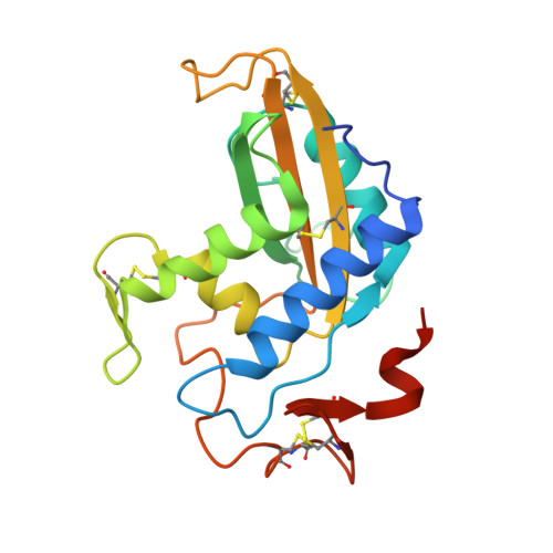
    Human glioma pathogenesis-related protein 1 (GLIPR1) is a membrane protein that is highly upregulated in brain cancers but is barely detectable in normal brain tissue. GLIPR1 is composed of a signal peptide that directs its secretion, a conserved cysteine-rich CAP (cysteine-rich secretory proteins, antigen 5 and pathogenesis-related 1 proteins) domain and a transmembrane domain. GLIPR1 is currently being investigated as a candidate for prostate cancer gene therapy and for glioblastoma targeted therapy. Crystal structures of a truncated soluble domain of the human GLIPR1 protein (sGLIPR1) solved by molecular replacement using a truncated polyalanine search model of the CAP domain of stecrisp, a snake-venom cysteine-rich secretory protein (CRISP), are presented. The correct molecular-replacement solution could only be obtained by removing all loops from the search model. The native structure was refined to 1.85 Å resolution and that of a Zn2+ complex was refined to 2.2 Å resolution. The latter structure revealed that the putative binding cavity coordinates Zn2+ similarly to snake-venom CRISPs, which are involved in Zn2+-dependent mechanisms of inflammatory modulation. Both sGLIPR1 structures have extensive flexible loop/turn regions and unique charge distributions that were not observed in any of the previously reported CAP protein structures. A model is also proposed for the structure of full-length membrane-bound GLIPR1.


  • Organizational Affiliation

    Department of Pathology and Microbiology, College of Medicine, Nebraska Medical Center, Omaha, NE 68198-6495, USA. oasojo@unmc.edu


Macromolecules
Find similar proteins by:  (by identity cutoff)  |  3D Structure
Entity ID: 1
MoleculeChains Sequence LengthOrganismDetailsImage
Glioma pathogenesis-related protein 1205Homo sapiensMutation(s): 0 
Gene Names: GLIPRGLIPR1RTVP1synthesized
UniProt & NIH Common Fund Data Resources
Find proteins for P48060 (Homo sapiens)
Explore P48060 
Go to UniProtKB:  P48060
PHAROS:  P48060
GTEx:  ENSG00000139278 
Entity Groups  
Sequence Clusters30% Identity50% Identity70% Identity90% Identity95% Identity100% Identity
UniProt GroupP48060
Sequence Annotations
Expand
  • Reference Sequence
Small Molecules
Ligands 1 Unique
IDChains Name / Formula / InChI Key2D Diagram3D Interactions
ZN
Query on ZN

Download Ideal Coordinates CCD File 
B [auth A]ZINC ION
Zn
PTFCDOFLOPIGGS-UHFFFAOYSA-N
Experimental Data & Validation

Experimental Data

  • Method: X-RAY DIFFRACTION
  • Resolution: 2.20 Å
  • R-Value Free: 0.192 
  • R-Value Work: 0.158 
  • R-Value Observed: 0.160 
  • Space Group: P 21 21 2
Unit Cell:
Length ( Å )Angle ( ˚ )
a = 85.92α = 90
b = 79.72β = 90
c = 38.76γ = 90
Software Package:
Software NamePurpose
PHENIXrefinement
PHASERphasing
REFMACrefinement
CrysalisProdata reduction
SCALAdata scaling
CrysalisProdata collection

Structure Validation

View Full Validation Report


Entry History 

Deposition Data

  • Released Date: 2011-10-05 
  • Deposition Author(s): Asojo, O.A.

Revision History  (Full details and data files)

  • Version 1.0: 2011-10-05
    Type: Initial release
  • Version 1.1: 2017-11-08
    Changes: Refinement description
  • Version 1.2: 2019-07-17
    Changes: Data collection, Refinement description
  • Version 1.3: 2023-09-13
    Changes: Data collection, Database references, Derived calculations, Refinement description