3PR2

Tryptophan synthase indoline quinonoid structure with F9 inhibitor in alpha site


Experimental Data Snapshot

  • Method: X-RAY DIFFRACTION
  • Resolution: 1.85 Å
  • R-Value Free: 0.227 
  • R-Value Work: 0.198 
  • R-Value Observed: 0.200 

wwPDB Validation   3D Report Full Report


Ligand Structure Quality Assessment 


This is version 1.4 of the entry. See complete history


Literature

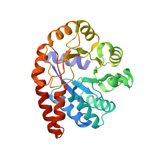
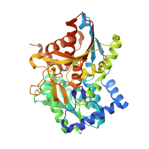
X-ray and NMR Crystallography in an Enzyme Active Site: The Indoline Quinonoid Intermediate in Tryptophan Synthase.

Lai, J.Niks, D.Wang, Y.Domratcheva, T.Barends, T.R.Schwarz, F.Olsen, R.A.Elliott, D.W.Fatmi, M.Q.Chang, C.E.Schlichting, I.Dunn, M.F.Mueller, L.J.

(2011) J Am Chem Soc 133: 4-7

  • DOI: https://doi.org/10.1021/ja106555c
  • Primary Citation of Related Structures:  
    3PR2

  • PubMed Abstract: 

    Chemical-level details such as protonation and hybridization state are critical for understanding enzyme mechanism and function. Even at high resolution, these details are difficult to determine by X-ray crystallography alone. The chemical shift in NMR spectroscopy, however, is an extremely sensitive probe of the chemical environment, making solid-state NMR spectroscopy and X-ray crystallography a powerful combination for defining chemically detailed three-dimensional structures. Here we adopted this combined approach to determine the chemically rich crystal structure of the indoline quinonoid intermediate in the pyridoxal-5'-phosphate-dependent enzyme tryptophan synthase under conditions of active catalysis. Models of the active site were developed using a synergistic approach in which the structure of this reactive substrate analogue was optimized using ab initio computational chemistry in the presence of side-chain residues fixed at their crystallographically determined coordinates. Various models of charge and protonation state for the substrate and nearby catalytic residues could be uniquely distinguished by their calculated effects on the chemical shifts measured at specifically (13)C- and (15)N-labeled positions on the substrate. Our model suggests the importance of an equilibrium between tautomeric forms of the substrate, with the protonation state of the major isomer directing the next catalytic step.


  • Organizational Affiliation

    Department of Chemistry, University of California, Riverside, California 92521, USA.


Macromolecules
Find similar proteins by:  (by identity cutoff)  |  3D Structure
Entity ID: 1
MoleculeChains Sequence LengthOrganismDetailsImage
Tryptophan synthase alpha chain266Salmonella enterica subsp. enterica serovar TyphimuriumMutation(s): 0 
Gene Names: STM1727trpA
EC: 4.2.1.20
UniProt
Find proteins for P00929 (Salmonella typhimurium (strain LT2 / SGSC1412 / ATCC 700720))
Explore P00929 
Go to UniProtKB:  P00929
Entity Groups  
Sequence Clusters30% Identity50% Identity70% Identity90% Identity95% Identity100% Identity
UniProt GroupP00929
Sequence Annotations
Expand
  • Reference Sequence
Find similar proteins by:  (by identity cutoff)  |  3D Structure
Entity ID: 2
MoleculeChains Sequence LengthOrganismDetailsImage
Tryptophan synthase beta chain391Salmonella enterica subsp. enterica serovar TyphimuriumMutation(s): 0 
Gene Names: STM1726trpB
EC: 4.2.1.20
UniProt
Find proteins for P0A2K1 (Salmonella typhimurium (strain LT2 / SGSC1412 / ATCC 700720))
Explore P0A2K1 
Go to UniProtKB:  P0A2K1
Entity Groups  
Sequence Clusters30% Identity50% Identity70% Identity90% Identity95% Identity100% Identity
UniProt GroupP0A2K1
Sequence Annotations
Expand
  • Reference Sequence
Small Molecules
Ligands 3 Unique
IDChains Name / Formula / InChI Key2D Diagram3D Interactions
7MN
Query on 7MN

Download Ideal Coordinates CCD File 
D [auth B](Z)-N-[(1E)-1-carboxy-2-(2,3-dihydro-1H-indol-1-yl)ethylidene]{3-hydroxy-2-methyl-5-[(phosphonooxy)methyl]pyridin-4(1H)-ylidene}methanaminium
C19 H23 N3 O7 P
MAJPVKFOYDGVKD-LNHPUNTFSA-O
F9F
Query on F9F

Download Ideal Coordinates CCD File 
C [auth A]2-({[4-(TRIFLUOROMETHOXY)PHENYL]SULFONYL}AMINO)ETHYL DIHYDROGEN PHOSPHATE
C9 H11 F3 N O7 P S
JDDKDMFCTOZVCJ-UHFFFAOYSA-N
CS
Query on CS

Download Ideal Coordinates CCD File 
E [auth B],
F [auth B],
G [auth B]
CESIUM ION
Cs
NCMHKCKGHRPLCM-UHFFFAOYSA-N
Experimental Data & Validation

Experimental Data

  • Method: X-RAY DIFFRACTION
  • Resolution: 1.85 Å
  • R-Value Free: 0.227 
  • R-Value Work: 0.198 
  • R-Value Observed: 0.200 
  • Space Group: C 1 2 1
Unit Cell:
Length ( Å )Angle ( ˚ )
a = 184.17α = 90
b = 60.62β = 94.62
c = 67.38γ = 90
Software Package:
Software NamePurpose
REFMACrefinement
CNSrefinement
MAR345data collection
XDSdata reduction
XSCALEdata scaling
CNSphasing

Structure Validation

View Full Validation Report


Ligand Structure Quality Assessment 


Entry History 

Revision History  (Full details and data files)

  • Version 1.0: 2011-02-09
    Type: Initial release
  • Version 1.1: 2011-07-13
    Changes: Version format compliance
  • Version 1.2: 2017-11-08
    Changes: Refinement description
  • Version 1.3: 2019-07-17
    Changes: Data collection, Refinement description
  • Version 1.4: 2024-02-21
    Changes: Data collection, Database references, Derived calculations