3PQ1

Crystal structure of human mitochondrial poly(A) polymerase (PAPD1)


Experimental Data Snapshot

  • Method: X-RAY DIFFRACTION
  • Resolution: 3.10 Å
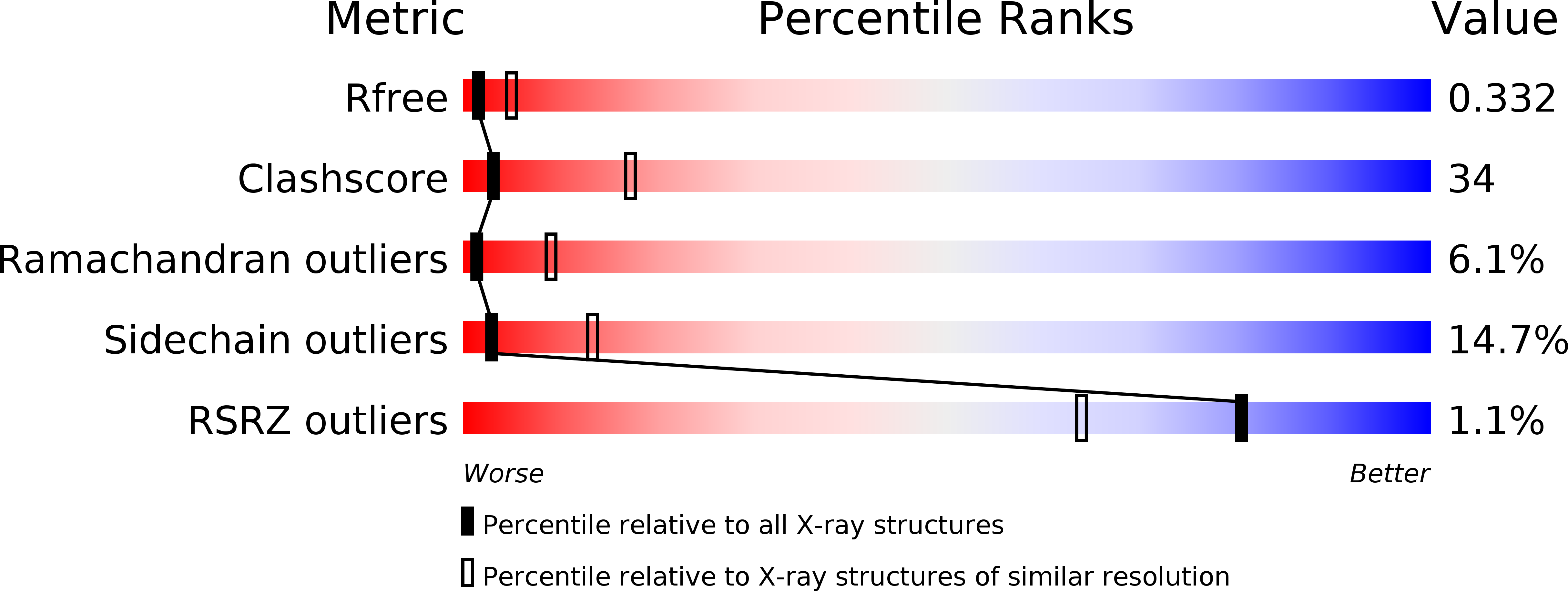
  • R-Value Free: 0.328 
  • R-Value Work: 0.247 
  • R-Value Observed: 0.251 

wwPDB Validation   3D Report Full Report


This is version 1.2 of the entry. See complete history


Literature

Structural basis for dimerization and activity of human PAPD1, a noncanonical poly(A) polymerase.

Bai, Y.Srivastava, S.K.Chang, J.H.Manley, J.L.Tong, L.

(2011) Mol Cell 41: 311-320

  • DOI: https://doi.org/10.1016/j.molcel.2011.01.013
  • Primary Citation of Related Structures:  
    3PQ1

  • PubMed Abstract: 

    Poly(A) polymerases (PAPs) are found in most living organisms and have important roles in RNA function and metabolism. Here, we report the crystal structure of human PAPD1, a noncanonical PAP that can polyadenylate RNAs in the mitochondria (also known as mtPAP) and oligouridylate histone mRNAs (TUTase1). The overall structure of the palm and fingers domains is similar to that in the canonical PAPs. The active site is located at the interface between the two domains, with a large pocket that can accommodate the substrates. The structure reveals the presence of a previously unrecognized domain in the N-terminal region of PAPD1, with a backbone fold that is similar to that of RNP-type RNA binding domains. This domain (named the RL domain), together with a β-arm insertion in the palm domain, contributes to dimerization of PAPD1. Surprisingly, our mutagenesis and biochemical studies show that dimerization is required for the catalytic activity of PAPD1.


  • Organizational Affiliation

    Department of Biological Sciences, Columbia University, New York, NY 10027, USA.


Macromolecules
Find similar proteins by:  (by identity cutoff)  |  3D Structure
Entity ID: 1
MoleculeChains Sequence LengthOrganismDetailsImage
Poly(A) RNA polymerase
A, B
464Homo sapiensMutation(s): 1 
Gene Names: MTPAPPAPD1
EC: 2.7.7.19
UniProt & NIH Common Fund Data Resources
Find proteins for Q9NVV4 (Homo sapiens)
Explore Q9NVV4 
Go to UniProtKB:  Q9NVV4
PHAROS:  Q9NVV4
GTEx:  ENSG00000107951 
Entity Groups  
Sequence Clusters30% Identity50% Identity70% Identity90% Identity95% Identity100% Identity
UniProt GroupQ9NVV4
Sequence Annotations
Expand
  • Reference Sequence
Small Molecules
Modified Residues  1 Unique
IDChains TypeFormula2D DiagramParent
MSE
Query on MSE
A, B
L-PEPTIDE LINKINGC5 H11 N O2 SeMET
Experimental Data & Validation

Experimental Data

  • Method: X-RAY DIFFRACTION
  • Resolution: 3.10 Å
  • R-Value Free: 0.328 
  • R-Value Work: 0.247 
  • R-Value Observed: 0.251 
  • Space Group: P 1 21 1
Unit Cell:
Length ( Å )Angle ( ˚ )
a = 68.68α = 90
b = 76.88β = 103.39
c = 87.63γ = 90
Software Package:
Software NamePurpose
ADSCdata collection
SOLVEphasing
PHENIXrefinement
DENZOdata reduction
SCALEPACKdata scaling

Structure Validation

View Full Validation Report


Entry History 

Deposition Data

Revision History  (Full details and data files)

  • Version 1.0: 2011-03-30
    Type: Initial release
  • Version 1.1: 2011-07-13
    Changes: Version format compliance
  • Version 1.2: 2017-08-02
    Changes: Refinement description, Source and taxonomy