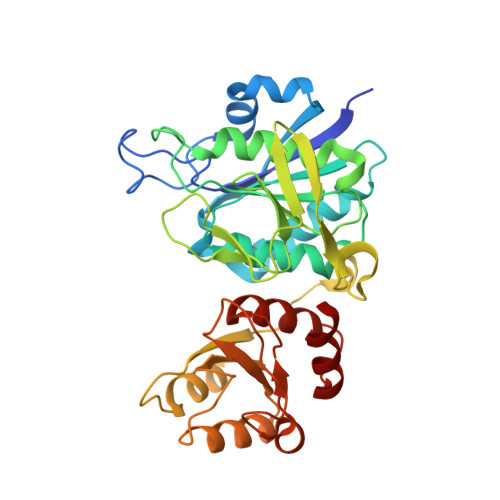
Structural characterization of Pseudomonas 7A glutaminase-asparaginase.
Lubkowski, J., Wlodawer, A., Ammon, H.L., Copeland, T.D., Swain, A.L.(1994) Biochemistry 33: 10257-10265
- PubMed: 8068664
- DOI: https://doi.org/10.1021/bi00200a005
- Primary Citation of Related Structures:
3PGA - PubMed Abstract:
The amino acid sequence and a 2-A-resolution crystallographic structure of Pseudomonas 7A glutaminase-asparaginase (PGA) have been determined. PGA, which belongs to the family of tetrameric bacterial amidohydrolases, deamidates glutamine and asparagine. The amino acid sequence of PGA has a high degree of similarity to the sequences of other members of the family. PGA has the same fold as other bacterial amidohydrolases, with the exception of the position of a 20-residue loop that forms part of the active site. In the PGA structure presented here, the active site loop is observed clearly in only one monomer, in an open position, with a conformation different from that observed for other amidohydrolases. In the other three monomers the loop is disordered and cannot be traced. This phenomenon is probably a direct consequence of a very low occupancy of product(s) of the enzymatic reaction bound in the active sites of PGA in these crystals. The active sites are composed of a rigid part and the flexible loop. The rigid part consists of the residues directly involved in the catalytic reaction as well as residues that assist in orienting the substrate. Two residues that are important for activity residue on the flexible loop. We suggest that the flexible loops actively participate in the transport of substrate and product molecules through the amidohydrolase active sites and participate in orienting the substrate molecules properly in relation to the catalytic residues.
Organizational Affiliation:
Macromolecular Structure Laboratory, NCI-Frederick Cancer Research and Development Center, Maryland 21702-1201.