3P72

structure of platelet Glycoprotein 1b alpha with a bound peptide inhibitor


Experimental Data Snapshot

  • Method: X-RAY DIFFRACTION
  • Resolution: 1.90 Å
  • R-Value Free: 0.286 
  • R-Value Work: 0.217 
  • R-Value Observed: 0.221 

wwPDB Validation   3D Report Full Report


This is version 1.3 of the entry. See complete history


Literature

Glycoprotein Ibalpha inhibitor complex structure reveals a combined steric and allosteric mechanism of von Willebrand factor antagonism.

McEwan, P.A.Andrews, R.K.Emsley, J.

(2009) Blood 114: 4883-4885

  • DOI: https://doi.org/10.1182/blood-2009-05-224170
  • Primary Citation of Related Structures:  
    3P72

  • PubMed Abstract: 

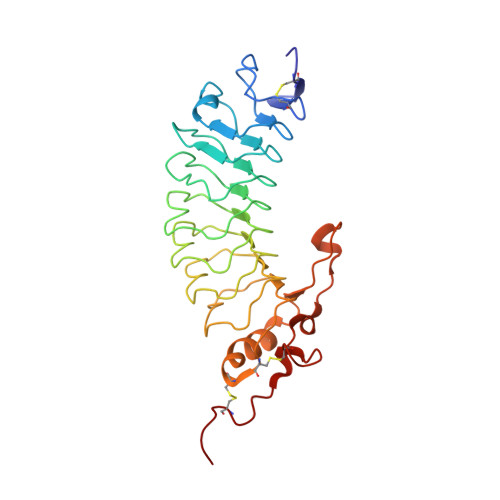
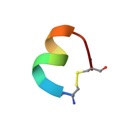
    Platelet glycoprotein Ibalpha (GpIbalpha) interactions with von Willebrand factor (VWF) are a critical early event in platelet adhesion, which contributes to hemostasis and thrombosis. Here we report the structure of a complex between GpIbalpha and a potent peptide inhibitor. The cyclic peptide (CTERMALHNLC) was isolated from a cysteine-constrained phage display library, and in the complex this forms one and a half turns of an amphipathic alpha-helix, the curvature of which facilitates contacts with the curved concave face of the GpIbalpha leucine-rich repeats. The peptide has only limited overlap with the VWF binding site. It effectively inhibits by stabilizing an alternative alpha-helical conformation of a regulatory loop that forms an extended beta-hairpin upon VWF binding. The structure defines a previously unrecognized binding site within GpIbalpha and represents a clear strategy for developing antiplatelet agents targeting the GpIbalpha-VWF interaction allosterically.


  • Organizational Affiliation

    Centre for Biomolecular Sciences, School of Pharmacy, University of Nottingham, Nottingham, United Kingdom.


Macromolecules
Find similar proteins by:  (by identity cutoff)  |  3D Structure
Entity ID: 1
MoleculeChains Sequence LengthOrganismDetailsImage
Platelet glycoprotein Ib alpha chain269Homo sapiensMutation(s): 2 
Gene Names: GP1BA
UniProt & NIH Common Fund Data Resources
Find proteins for P07359 (Homo sapiens)
Explore P07359 
Go to UniProtKB:  P07359
PHAROS:  P07359
GTEx:  ENSG00000185245 
Entity Groups  
Sequence Clusters30% Identity50% Identity70% Identity90% Identity95% Identity100% Identity
UniProt GroupP07359
Sequence Annotations
Expand
  • Reference Sequence

Find similar proteins by:  Sequence   |   3D Structure  

Entity ID: 2
MoleculeChains Sequence LengthOrganismDetailsImage
OS1 peptide11N/AMutation(s): 0 
Entity Groups  
Sequence Clusters30% Identity50% Identity70% Identity90% Identity95% Identity100% Identity
Sequence Annotations
Expand
  • Reference Sequence
Small Molecules
Ligands 1 Unique
IDChains Name / Formula / InChI Key2D Diagram3D Interactions
CL
Query on CL

Download Ideal Coordinates CCD File 
C [auth A]CHLORIDE ION
Cl
VEXZGXHMUGYJMC-UHFFFAOYSA-M
Biologically Interesting Molecules (External Reference) 1 Unique
Experimental Data & Validation

Experimental Data

  • Method: X-RAY DIFFRACTION
  • Resolution: 1.90 Å
  • R-Value Free: 0.286 
  • R-Value Work: 0.217 
  • R-Value Observed: 0.221 
  • Space Group: P 31 2 1
Unit Cell:
Length ( Å )Angle ( ˚ )
a = 58.817α = 90
b = 58.817β = 90
c = 163.027γ = 120
Software Package:
Software NamePurpose
DNAdata collection
PHASERphasing
REFMACrefinement
MOSFLMdata reduction
SCALAdata scaling

Structure Validation

View Full Validation Report


Entry History 

Deposition Data

Revision History  (Full details and data files)

  • Version 1.0: 2010-11-24
    Type: Initial release
  • Version 1.1: 2011-07-13
    Changes: Version format compliance
  • Version 1.2: 2012-12-12
    Changes: Other
  • Version 1.3: 2018-10-10
    Changes: Data collection, Database references, Structure summary