3P42

Structure of GfcC (YmcB), protein encoded by the E. coli group 4 capsule operon


Experimental Data Snapshot

  • Method: X-RAY DIFFRACTION
  • Resolution: 1.91 Å
  • R-Value Free: 0.217 
  • R-Value Work: 0.173 
  • R-Value Observed: 0.175 

wwPDB Validation   3D Report Full Report


This is version 1.2 of the entry. See complete history


Literature

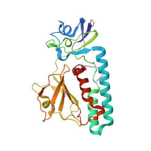
The Crystal Structure of Escherichia coli Group 4 Capsule Protein GfcC Reveals a Domain Organization Resembling That of Wza.

Sathiyamoorthy, K.Mills, E.Franzmann, T.M.Rosenshine, I.Saper, M.A.

(2011) Biochemistry 50: 5465-5476

  • DOI: https://doi.org/10.1021/bi101869h
  • Primary Citation of Related Structures:  
    3P42

  • PubMed Abstract: 

    We report the 1.9 Å resolution crystal structure of enteropathogenic Escherichia coli GfcC, a periplasmic protein encoded by the gfc operon, which is essential for assembly of group 4 polysaccharide capsule (O-antigen capsule). Presumed gene orthologs of gfcC are present in capsule-encoding regions of at least 29 genera of Gram-negative bacteria. GfcC, a member of the DUF1017 family, is comprised of tandem β-grasp (ubiquitin-like) domains (D2 and D3) and a carboxyl-terminal amphipathic helix, a domain arrangement reminiscent of that of Wza that forms an exit pore for group 1 capsule export. Unlike the membrane-spanning C-terminal helix from Wza, the GfcC C-terminal helix packs against D3. Previously unobserved in a β-grasp domain structure is a 48-residue helical hairpin insert in D2 that binds to D3, constraining its position and sequestering the carboxyl-terminal amphipathic helix. A centrally located and invariant Arg115 not only is essential for proper localization but also forms one of two mostly conserved pockets. Finally, we draw analogies between a GfcC protein fused to an outer membrane β-barrel pore in some species and fusion proteins necessary for secreting biofilm-forming exopolysaccharides.


  • Organizational Affiliation

    Program in Biophysics, University of Michigan, Ann Arbor, Michigan 48109-1055, USA.


Macromolecules
Find similar proteins by:  (by identity cutoff)  |  3D Structure
Entity ID: 1
MoleculeChains Sequence LengthOrganismDetailsImage
Predicted protein
A, B, C, D
236Escherichia coli O127:H6 str. E2348/69Mutation(s): 0 
Gene Names: E2348C_0970E2348_C_0970gfcC
UniProt
Find proteins for B7UN63 (Escherichia coli O127:H6 (strain E2348/69 / EPEC))
Explore B7UN63 
Go to UniProtKB:  B7UN63
Entity Groups  
Sequence Clusters30% Identity50% Identity70% Identity90% Identity95% Identity100% Identity
UniProt GroupB7UN63
Sequence Annotations
Expand
  • Reference Sequence
Small Molecules
Modified Residues  1 Unique
IDChains TypeFormula2D DiagramParent
MSE
Query on MSE
A, B, C, D
L-PEPTIDE LINKINGC5 H11 N O2 SeMET
Experimental Data & Validation

Experimental Data

  • Method: X-RAY DIFFRACTION
  • Resolution: 1.91 Å
  • R-Value Free: 0.217 
  • R-Value Work: 0.173 
  • R-Value Observed: 0.175 
  • Space Group: P 1 21 1
Unit Cell:
Length ( Å )Angle ( ˚ )
a = 68.831α = 90
b = 99.98β = 91.74
c = 69.02γ = 90
Software Package:
Software NamePurpose
MAR345data collection
PHASERphasing
PHENIXrefinement
HKL-2000data reduction
HKL-2000data scaling

Structure Validation

View Full Validation Report


Entry History 

Deposition Data

Revision History  (Full details and data files)

  • Version 1.0: 2011-04-06
    Type: Initial release
  • Version 1.1: 2011-07-13
    Changes: Version format compliance
  • Version 1.2: 2017-11-08
    Changes: Refinement description