3P12

Crystal Structure of D-ribose Pyranase Sa240


Experimental Data Snapshot

  • Method: X-RAY DIFFRACTION
  • Resolution: 2.35 Å
  • R-Value Free: 0.252 
  • R-Value Work: 0.217 
  • R-Value Observed: 0.219 

wwPDB Validation   3D Report Full Report


This is version 1.2 of the entry. See complete history


Literature

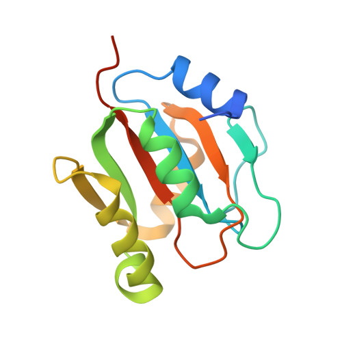
Crystal structure of Sa240: A ribose pyranase homolog with partial active site from Staphylococcus aureus

Wang, L.Wu, M.Zang, J.

(2011) J Struct Biol 174: 413-419

  • DOI: https://doi.org/10.1016/j.jsb.2011.01.007
  • Primary Citation of Related Structures:  
    3P12, 3P13

  • PubMed Abstract: 

    Ribose is transported into cells in its pyranose form and must be rearranged to its furanose form for further utilization. Ribose pyranase RbsD catalyzes the conversion of ribose from the pyranose to furanose form. This is the key step for substrate supply to ribokinase RbsK, which converts ribose to ribose-5-phosphate for further metabolism. Sequence analysis indicated Sa240 from Staphylococcus aureus was a ribose pyranase homolog. Here we showed that Sa240 formed dimeric structure both in solution and in crystal. S240-ribose complex structure showed a ribose binding site formed by an incomplete active site compared with RbsD. Because the catalytic activity of ribose pyranase depends on its oligomeric state, we propose Sa240 is catalytically inactive in its dimeric structure.


  • Organizational Affiliation

    School of Life Sciences, University of Science and Technology of China, 96 Jinzhai Road, Hefei, Anhui 230026, People's Republic of China.


Macromolecules
Find similar proteins by:  (by identity cutoff)  |  3D Structure
Entity ID: 1
MoleculeChains Sequence LengthOrganismDetailsImage
D-ribose pyranase
A, B, C, D
144Staphylococcus aureus subsp. aureus NCTC 8325Mutation(s): 0 
Gene Names: rbsDSAOUHSC_00240
EC: 5.5.1
UniProt
Find proteins for Q2G1A5 (Staphylococcus aureus (strain NCTC 8325 / PS 47))
Explore Q2G1A5 
Go to UniProtKB:  Q2G1A5
Entity Groups  
Sequence Clusters30% Identity50% Identity70% Identity90% Identity95% Identity100% Identity
UniProt GroupQ2G1A5
Sequence Annotations
Expand
  • Reference Sequence
Experimental Data & Validation

Experimental Data

  • Method: X-RAY DIFFRACTION
  • Resolution: 2.35 Å
  • R-Value Free: 0.252 
  • R-Value Work: 0.217 
  • R-Value Observed: 0.219 
  • Space Group: P 65 2 2
Unit Cell:
Length ( Å )Angle ( ˚ )
a = 122.126α = 90
b = 122.126β = 90
c = 175.861γ = 120
Software Package:
Software NamePurpose
MAR345dtbdata collection
PHENIXmodel building
REFMACrefinement
HKL-2000data reduction
HKL-2000data scaling
PHENIXphasing

Structure Validation

View Full Validation Report


Entry History 

Deposition Data

Revision History  (Full details and data files)

  • Version 1.0: 2011-05-25
    Type: Initial release
  • Version 1.1: 2011-07-13
    Changes: Version format compliance
  • Version 1.2: 2024-03-20
    Changes: Data collection, Database references, Derived calculations