3NVU

Modulating Heme Redox Potential Through Protein-Induced Porphyrin Distortion


Experimental Data Snapshot

  • Method: X-RAY DIFFRACTION
  • Resolution: 2.04 Å
  • R-Value Free: 0.234 
  • R-Value Work: 0.189 
  • R-Value Observed: 0.192 

wwPDB Validation   3D Report Full Report


Ligand Structure Quality Assessment 


This is version 1.2 of the entry. See complete history


Literature

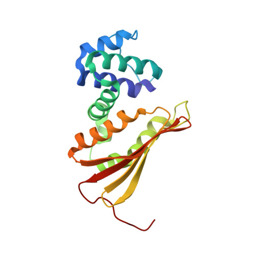
Modulating heme redox potential through protein-induced porphyrin distortion.

Olea, C.Kuriyan, J.Marletta, M.A.

(2010) J Am Chem Soc 132: 12794-12795

  • DOI: https://doi.org/10.1021/ja106252b
  • Primary Citation of Related Structures:  
    3NVR, 3NVU

  • PubMed Abstract: 

    Hemoproteins are ubiquitous in biology and are commonly involved in critical processes such as electron transfer, oxidative phosphorylation, and signal transduction. Both the protein environment and the heme cofactor contribute to generate the range of chemical properties needed for these diverse functions. Using the heme nitric oxide/oxygen binding (H-NOX) protein from the thermophilic bacterium Thermoanaerobacter tengcongensis, we have shown that heme electronic properties can be modulated by porphyrin distortion within the same protein scaffold without changing the heme ligation state or heme environment. The degree of heme distortion was found to be directly correlated to the electron density at the heme iron, as evidenced by dramatic changes in the heme redox potential and pK(a) of the distal ligand ((-)OH vs H(2)O). Protein-induced porphyrin distortion represents a new strategy to rationally tune the electronic properties of protein-bound porphyrins and could be used to engineer proteins with desired reactivity or functionality.


  • Organizational Affiliation

    Department of Molecular and Cell Biology, California Institute for Quantitative Biosciences, and Division of Physical Biosciences, University of California, Berkeley, Berkeley, California 94720-3220, USA.


Macromolecules
Find similar proteins by:  (by identity cutoff)  |  3D Structure
Entity ID: 1
MoleculeChains Sequence LengthOrganismDetailsImage
Methyl-accepting chemotaxis protein
A, B
188Caldanaerobacter subterraneus subsp. tengcongensisMutation(s): 2 
Gene Names: Tar4TTE0680
UniProt
Find proteins for Q8RBX6 (Caldanaerobacter subterraneus subsp. tengcongensis (strain DSM 15242 / JCM 11007 / NBRC 100824 / MB4))
Explore Q8RBX6 
Go to UniProtKB:  Q8RBX6
Entity Groups  
Sequence Clusters30% Identity50% Identity70% Identity90% Identity95% Identity100% Identity
UniProt GroupQ8RBX6
Sequence Annotations
Expand
  • Reference Sequence
Experimental Data & Validation

Experimental Data

  • Method: X-RAY DIFFRACTION
  • Resolution: 2.04 Å
  • R-Value Free: 0.234 
  • R-Value Work: 0.189 
  • R-Value Observed: 0.192 
  • Space Group: C 1 2 1
Unit Cell:
Length ( Å )Angle ( ˚ )
a = 127.158α = 90
b = 79.828β = 102.25
c = 43.322γ = 90
Software Package:
Software NamePurpose
ADSCdata collection
PHASERphasing
PHENIXrefinement
HKL-2000data reduction
HKL-2000data scaling

Structure Validation

View Full Validation Report


Ligand Structure Quality Assessment 


Entry History 

Deposition Data

Revision History  (Full details and data files)

  • Version 1.0: 2010-09-08
    Type: Initial release
  • Version 1.1: 2011-07-13
    Changes: Version format compliance
  • Version 1.2: 2024-02-21
    Changes: Data collection, Database references, Derived calculations