3MJ4

Crystal structure of UDP-galactopyranose mutase in complex with phosphonate analog of UDP-galactopyranose


Experimental Data Snapshot

  • Method: X-RAY DIFFRACTION
  • Resolution: 2.65 Å
  • R-Value Free: 0.262 
  • R-Value Work: 0.207 
  • R-Value Observed: 0.210 

wwPDB Validation   3D Report Full Report


Ligand Structure Quality Assessment 


This is version 1.4 of the entry. See complete history


Literature

Chemoenzymatic Synthesis, Inhibition Studies, and X-ray Crystallographic Analysis of the Phosphono Analog of UDP-Galp as an Inhibitor and Mechanistic Probe for UDP-Galactopyranose Mutase.

Partha, S.K.Sadeghi-Khomami, A.Slowski, K.Kotake, T.Thomas, N.R.Jakeman, D.L.Sanders, D.A.

(2010) J Mol Biol 403: 578-590

  • DOI: https://doi.org/10.1016/j.jmb.2010.08.053
  • Primary Citation of Related Structures:  
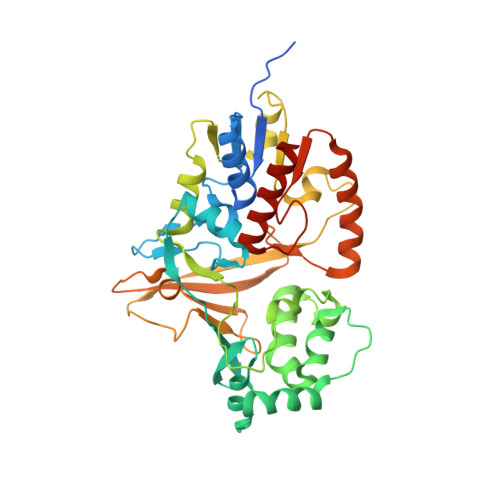
    3MJ4

  • PubMed Abstract: 

    UDP (uridine diphosphate) galactopyranose mutase (UGM) is involved in the cell wall biosynthesis of many pathogenic microorganisms. UGM catalyzes the reversible conversion of UDP-α-D-galactopyranose into UDP-α-D-galactofuranose, with the latter being the precursor of galactofuranose (Galf) residues in cell walls. Glycoconjugates of Galf are essential components in the cell wall of various pathogenic bacteria, including Mycobacterium tuberculosis, the causative agent of tuberculosis. The absence of Galf in humans and its bacterial requirement make UGM a potential target for developing novel antibacterial agents. In this article, we report the synthesis, inhibitory activity, and X-ray crystallographic studies of UDP-phosphono-galactopyranose, a nonhydrolyzable C-glycosidic phosphonate. This is the first report on the synthesis of a phosphonate analog of UDP-α-D-galactopyranose by a chemoenzymatic phosphoryl coupling method. The phosphonate was evaluated against three bacterial UGMs and showed only moderate inhibition. We determined the crystal structure of the phosphonate analog bound to Deinococcus radiodurans UGM at 2.6 Å resolution. The phosphonate analog is bound in a novel conformation not observed in UGM-substrate complex structures or in other enzyme-sugar nucleotide phosphonate complexes. This complex structure provides a structural basis for the observed micromolar inhibition towards UGM. Steric clashes, loss of electrostatic stabilization between an active-site arginine (Arg305) and the phosphonate analog, and a 180° flip of the hexose moiety account for the differences in the binding orientations of the isosteric phosphonate analog and the physiological substrate. This provides new insight into the ability of a sugar-nucleotide-binding enzyme to orient a substrate analog in an unexpected geometry and should be taken into consideration in designing such enzyme inhibitors.


  • Organizational Affiliation

    Department of Chemistry, University of Saskatchewan, Saskatoon, Saskatchewan, Canada.


Macromolecules
Find similar proteins by:  (by identity cutoff)  |  3D Structure
Entity ID: 1
MoleculeChains Sequence LengthOrganismDetailsImage
UDP-galactopyranose mutase
A, B, C, D, E
A, B, C, D, E, F, G, H, I, J
397Deinococcus radiodurans R1 = ATCC 13939 = DSM 20539Mutation(s): 0 
Gene Names: DR_A0367DR_AO367
EC: 5.4.99.9
UniProt
Find proteins for Q9RYF1 (Deinococcus radiodurans (strain ATCC 13939 / DSM 20539 / JCM 16871 / CCUG 27074 / LMG 4051 / NBRC 15346 / NCIMB 9279 / VKM B-1422 / R1))
Explore Q9RYF1 
Go to UniProtKB:  Q9RYF1
Entity Groups  
Sequence Clusters30% Identity50% Identity70% Identity90% Identity95% Identity100% Identity
UniProt GroupQ9RYF1
Sequence Annotations
Expand
  • Reference Sequence
Small Molecules
Ligands 5 Unique
IDChains Name / Formula / InChI Key2D Diagram3D Interactions
FAD
Query on FAD

Download Ideal Coordinates CCD File 
AA [auth E]
BB [auth J]
GA [auth F]
K [auth A]
LA [auth G]
AA [auth E],
BB [auth J],
GA [auth F],
K [auth A],
LA [auth G],
P [auth B],
QA [auth H],
S [auth C],
UA [auth I],
X [auth D]
FLAVIN-ADENINE DINUCLEOTIDE
C27 H33 N9 O15 P2
VWWQXMAJTJZDQX-UYBVJOGSSA-N
URM
Query on URM

Download Ideal Coordinates CCD File 
W [auth D](((2S,3R,4S,5R,6R)-3,4,5-trihydroxy-6-(hydroxymethyl)tetrahydro-2H-pyran-2-yl)methyl)phosphonic (((2R,3S,4R,5R)-5-(2,4-dioxo-3,4-dihydropyrimidin-1(2H)-yl)-3,4-dihydroxytetrahydrofuran-2-yl)methyl phosphoric) anhydride
C16 H26 N2 O16 P2
WUPLBUVQIJIOHV-PPSAJGQHSA-N
UDP
Query on UDP

Download Ideal Coordinates CCD File 
BA [auth E]
CB [auth J]
HA [auth F]
L [auth A]
MA [auth G]
BA [auth E],
CB [auth J],
HA [auth F],
L [auth A],
MA [auth G],
Q [auth B],
RA [auth H],
T [auth C],
WA [auth I]
URIDINE-5'-DIPHOSPHATE
C9 H14 N2 O12 P2
XCCTYIAWTASOJW-XVFCMESISA-N
XYL
Query on XYL

Download Ideal Coordinates CCD File 
JA [auth F],
VA [auth I]
Xylitol
C5 H12 O5
HEBKCHPVOIAQTA-SCDXWVJYSA-N
GOL
Query on GOL

Download Ideal Coordinates CCD File 
AB [auth I]
CA [auth E]
DA [auth E]
DB [auth J]
EA [auth E]
AB [auth I],
CA [auth E],
DA [auth E],
DB [auth J],
EA [auth E],
EB [auth J],
FA [auth E],
IA [auth F],
KA [auth F],
M [auth A],
N [auth A],
NA [auth G],
O [auth A],
OA [auth G],
PA [auth G],
R [auth B],
SA [auth H],
TA [auth H],
U [auth C],
V [auth C],
XA [auth I],
Y [auth D],
YA [auth I],
Z [auth D],
ZA [auth I]
GLYCEROL
C3 H8 O3
PEDCQBHIVMGVHV-UHFFFAOYSA-N
Experimental Data & Validation

Experimental Data

  • Method: X-RAY DIFFRACTION
  • Resolution: 2.65 Å
  • R-Value Free: 0.262 
  • R-Value Work: 0.207 
  • R-Value Observed: 0.210 
  • Space Group: P 21 21 21
Unit Cell:
Length ( Å )Angle ( ˚ )
a = 134.149α = 90
b = 175.715β = 90
c = 223.99γ = 90
Software Package:
Software NamePurpose
MOLREPphasing
PHENIXrefinement
MxDCdata collection
d*TREKdata reduction
d*TREKdata scaling

Structure Validation

View Full Validation Report


Ligand Structure Quality Assessment 


Entry History 

Deposition Data

Revision History  (Full details and data files)

  • Version 1.0: 2010-10-20
    Type: Initial release
  • Version 1.1: 2011-07-13
    Changes: Version format compliance
  • Version 1.2: 2017-11-08
    Changes: Advisory, Refinement description
  • Version 1.3: 2020-07-29
    Type: Remediation
    Reason: Carbohydrate remediation
    Changes: Derived calculations, Structure summary
  • Version 1.4: 2023-09-06
    Changes: Advisory, Data collection, Database references, Refinement description, Structure summary