3LVW

Glutathione-inhibited ScGCL


Experimental Data Snapshot

  • Method: X-RAY DIFFRACTION
  • Resolution: 2.50 Å
  • R-Value Free: 0.251 
  • R-Value Work: 0.199 
  • R-Value Observed: 0.202 

wwPDB Validation   3D Report Full Report


Ligand Structure Quality Assessment 


This is version 1.2 of the entry. See complete history


Literature

Structural basis for feedback and pharmacological inhibition of Saccharomyces cerevisiae glutamate cysteine ligase.

Biterova, E.I.Barycki, J.J.

(2010) J Biol Chem 285: 14459-14466

  • DOI: https://doi.org/10.1074/jbc.M110.104802
  • Primary Citation of Related Structures:  
    3LVV, 3LVW

  • PubMed Abstract: 

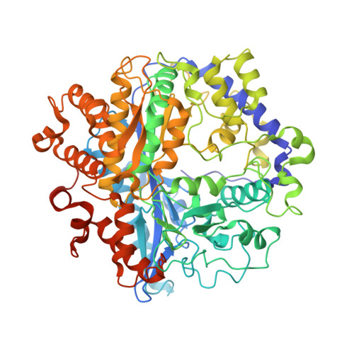
    Structural characterization of glutamate cysteine ligase (GCL), the enzyme that catalyzes the initial, rate-limiting step in glutathione biosynthesis, has revealed many of the molecular details of substrate recognition. To further delineate the mechanistic details of this critical enzyme, we have determined the structures of two inhibited forms of Saccharomyces cerevisiae GCL (ScGCL), which shares significant sequence identity with the human enzyme. In vivo, GCL activity is feedback regulated by glutathione. Examination of the structure of ScGCL-glutathione complex (2.5 A; R = 19.9%, R(free) = 25.1%) indicates that the inhibitor occupies both the glutamate- and the presumed cysteine-binding site and disrupts the previously observed Mg(2+) coordination in the ATP-binding site. l-Buthionine-S-sulfoximine (BSO) is a mechanism-based inhibitor of GCL and has been used extensively to deplete glutathione in cell culture and in vivo model systems. Inspection of the ScGCL-BSO structure (2.2 A; R = 18.1%, R(free) = 23.9%) confirms that BSO is phosphorylated on the sulfoximine nitrogen to generate the inhibitory species and reveals contacts that likely contribute to transition state stabilization. Overall, these structures advance our understanding of the molecular regulation of this critical enzyme and provide additional details of the catalytic mechanism of the enzyme.


  • Organizational Affiliation

    Department of Biochemistry and the Redox Biology Center, University of Nebraska, Lincoln, Nebraska 68588, USA.


Macromolecules
Find similar proteins by:  (by identity cutoff)  |  3D Structure
Entity ID: 1
MoleculeChains Sequence LengthOrganismDetailsImage
Glutamate--cysteine ligase692Saccharomyces cerevisiaeMutation(s): 0 
Gene Names: GSH1J0832YJL101C
EC: 6.3.2.2
UniProt
Find proteins for P32477 (Saccharomyces cerevisiae (strain ATCC 204508 / S288c))
Explore P32477 
Go to UniProtKB:  P32477
Entity Groups  
Sequence Clusters30% Identity50% Identity70% Identity90% Identity95% Identity100% Identity
UniProt GroupP32477
Sequence Annotations
Expand
  • Reference Sequence
Small Molecules
Binding Affinity Annotations 
IDSourceBinding Affinity
GSH PDBBind:  3LVW Ki: 2.12e+6 (nM) from 1 assay(s)
Binding MOAD:  3LVW Ki: 2.12e+6 (nM) from 1 assay(s)
Experimental Data & Validation

Experimental Data

  • Method: X-RAY DIFFRACTION
  • Resolution: 2.50 Å
  • R-Value Free: 0.251 
  • R-Value Work: 0.199 
  • R-Value Observed: 0.202 
  • Space Group: P 43 21 2
Unit Cell:
Length ( Å )Angle ( ˚ )
a = 118.127α = 90
b = 118.127β = 90
c = 165.799γ = 90
Software Package:
Software NamePurpose
CrystalCleardata collection
PHENIXmodel building
REFMACrefinement
HKL-2000data reduction
HKL-2000data scaling
PHENIXphasing

Structure Validation

View Full Validation Report


Ligand Structure Quality Assessment 


Entry History 

Deposition Data

Revision History  (Full details and data files)

  • Version 1.0: 2010-03-16
    Type: Initial release
  • Version 1.1: 2011-07-13
    Changes: Version format compliance
  • Version 1.2: 2023-09-06
    Changes: Data collection, Database references, Derived calculations, Refinement description