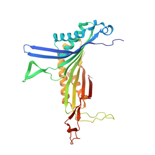
3LBG

Urate oxidase complexed with 8-thio xanthine


Experimental Data Snapshot

  • Method: X-RAY DIFFRACTION
  • Resolution: 1.50 Å
  • R-Value Free: 0.241 
  • R-Value Work: 0.201 
  • R-Value Observed: 0.202 

wwPDB Validation   3D Report Full Report


Ligand Structure Quality Assessment 


This is version 1.3 of the entry. See complete history


Literature

Near-atomic resolution structures of urate oxidase complexed with its substrate and analogues: the protonation state of the ligand.

Gabison, L.Chiadmi, M.El Hajji, M.Castro, B.Colloc'h, N.Prange, T.

(2010) Acta Crystallogr D Biol Crystallogr 66: 714-724

  • DOI: https://doi.org/10.1107/S090744491001142X
  • Primary Citation of Related Structures:  
    3L8W, 3L9G, 3LBG, 3LD4

  • PubMed Abstract: 

    Urate oxidase (uricase; EC 1.7.3.3; UOX) from Aspergillus flavus catalyzes the oxidation of uric acid in the presence of molecular oxygen to 5-hydroxyisourate in the degradation cascade of purines; intriguingly, catalysis proceeds using neither a metal ion (Fe, Cu etc.) nor a redox cofactor. UOX is a tetrameric enzyme with four active sites located at the interface of two subunits; its structure was refined at atomic resolution (1 A) using new crystal data in the presence of xanthine and at near-atomic resolution (1.3-1.7 A) in complexes with the natural substrate (urate) and two inhibitors: 8-nitroxanthine and 8-thiouric acid. Three new features of the structural and mechanistic behaviour of the enzyme were addressed. Firstly, the high resolution of the UOX-xanthine structure allowed the solution of an old structural problem at a contact zone within the tetramer; secondly, the protonation state of the substrate was determined from both a halochromic inhibitor complex (UOX-8-nitroxanthine) and from the H-atom distribution in the active site, using the structures of the UOX-xanthine and the UOX-uric acid complexes; and thirdly, it was possible to extend the general base system, characterized by the conserved catalytic triad Thr-Lys-His, to a large water network that is able to buffer and shuttle protons back and forth between the substrate and the peroxo hole along the reaction pathway.


  • Organizational Affiliation

    Laboratoire de Cristallographie et RMN Biologiques, UMR 8015 CNRS, France.


Macromolecules
Find similar proteins by:  (by identity cutoff)  |  3D Structure
Entity ID: 1
MoleculeChains Sequence LengthOrganismDetailsImage
Uricase296Aspergillus flavusMutation(s): 1 
EC: 1.7.3.3
UniProt
Find proteins for Q00511 (Aspergillus flavus)
Explore Q00511 
Go to UniProtKB:  Q00511
Entity Groups  
Sequence Clusters30% Identity50% Identity70% Identity90% Identity95% Identity100% Identity
UniProt GroupQ00511
Sequence Annotations
Expand
  • Reference Sequence
Small Molecules
Ligands 4 Unique
IDChains Name / Formula / InChI Key2D Diagram3D Interactions
8TX
Query on 8TX

Download Ideal Coordinates CCD File 
B [auth A],
C [auth A]
8-thioxo-3,7,8,9-tetrahydro-1H-purine-2,6-dione
C5 H4 N4 O2 S
KFQXXACDWBTCDT-UHFFFAOYSA-N
TRS
Query on TRS

Download Ideal Coordinates CCD File 
D [auth A]2-AMINO-2-HYDROXYMETHYL-PROPANE-1,3-DIOL
C4 H12 N O3
LENZDBCJOHFCAS-UHFFFAOYSA-O
EDO
Query on EDO

Download Ideal Coordinates CCD File 
E [auth A],
F [auth A],
G [auth A],
H [auth A]
1,2-ETHANEDIOL
C2 H6 O2
LYCAIKOWRPUZTN-UHFFFAOYSA-N
NA
Query on NA

Download Ideal Coordinates CCD File 
I [auth A]SODIUM ION
Na
FKNQFGJONOIPTF-UHFFFAOYSA-N
Experimental Data & Validation

Experimental Data

  • Method: X-RAY DIFFRACTION
  • Resolution: 1.50 Å
  • R-Value Free: 0.241 
  • R-Value Work: 0.201 
  • R-Value Observed: 0.202 
  • Space Group: I 2 2 2
Unit Cell:
Length ( Å )Angle ( ˚ )
a = 79.637α = 90
b = 95.471β = 90
c = 104.61γ = 90
Software Package:
Software NamePurpose
MxCuBEdata collection
SHELXmodel building
SHELXL-97refinement
MOSFLMdata reduction
SCALAdata scaling
SHELXphasing

Structure Validation

View Full Validation Report


Ligand Structure Quality Assessment 


Entry History 

Deposition Data

Revision History  (Full details and data files)

  • Version 1.0: 2010-06-02
    Type: Initial release
  • Version 1.1: 2011-07-13
    Changes: Version format compliance
  • Version 1.2: 2012-04-04
    Changes: Database references
  • Version 1.3: 2023-11-01
    Changes: Data collection, Database references, Derived calculations, Refinement description