3LAT

Crystal structure of Staphylococcus peptidoglycan hydrolase AmiE


Experimental Data Snapshot

  • Method: X-RAY DIFFRACTION
  • Resolution: 1.70 Å
  • R-Value Free: 0.165 
  • R-Value Work: 0.143 
  • R-Value Observed: 0.144 

wwPDB Validation   3D Report Full Report


This is version 1.2 of the entry. See complete history


Literature

Structural basis of cell wall cleavage by a staphylococcal autolysin

Zoll, S.Patzold, B.Schlag, M.Gotz, F.Kalbacher, H.Stehle, T.

(2010) PLoS Pathog 6: e1000807-e1000807

  • DOI: https://doi.org/10.1371/journal.ppat.1000807
  • Primary Citation of Related Structures:  
    3LAT

  • PubMed Abstract: 

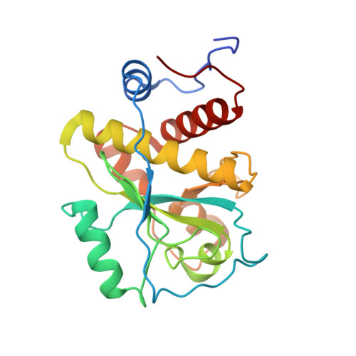
    The major autolysins (Atl) of Staphylococcus epidermidis and S. aureus play an important role in cell separation, and their mutants are also attenuated in virulence. Therefore, autolysins represent a promising target for the development of new types of antibiotics. Here, we report the high-resolution structure of the catalytically active amidase domain AmiE (amidase S. epidermidis) from the major autolysin of S. epidermidis. This is the first protein structure with an amidase-like fold from a bacterium with a gram-positive cell wall architecture. AmiE adopts a globular fold, with several alpha-helices surrounding a central beta-sheet. Sequence comparison reveals a cluster of conserved amino acids that define a putative binding site with a buried zinc ion. Mutations of key residues in the putative active site result in loss of activity, enabling us to propose a catalytic mechanism. We also identified and synthesized muramyltripeptide, the minimal peptidoglycan fragment that can be used as a substrate by the enzyme. Molecular docking and digestion assays with muramyltripeptide derivatives allow us to identify key determinants of ligand binding. This results in a plausible model of interaction of this ligand not only for AmiE, but also for other PGN-hydrolases that share the same fold. As AmiE active-site mutations also show a severe growth defect, our findings provide an excellent platform for the design of specific inhibitors that target staphylococcal cell separation and can thereby prevent growth of this pathogen.


  • Organizational Affiliation

    Interfaculty Institute for Biochemistry, University of Tübingen, Tübingen, Germany.


Macromolecules
Find similar proteins by:  (by identity cutoff)  |  3D Structure
Entity ID: 1
MoleculeChains Sequence LengthOrganismDetailsImage
Bifunctional autolysin
A, B
213Staphylococcus epidermidisMutation(s): 0 
Gene Names: atlE
EC: 3.5.1.28
UniProt
Find proteins for O33635 (Staphylococcus epidermidis)
Explore O33635 
Go to UniProtKB:  O33635
Entity Groups  
Sequence Clusters30% Identity50% Identity70% Identity90% Identity95% Identity100% Identity
UniProt GroupO33635
Sequence Annotations
Expand
  • Reference Sequence
Small Molecules
Ligands 4 Unique
IDChains Name / Formula / InChI Key2D Diagram3D Interactions
BU1
Query on BU1

Download Ideal Coordinates CCD File 
K [auth A],
T [auth B],
U [auth B]
1,4-BUTANEDIOL
C4 H10 O2
WERYXYBDKMZEQL-UHFFFAOYSA-N
IMD
Query on IMD

Download Ideal Coordinates CCD File 
C [auth A],
L [auth B]
IMIDAZOLE
C3 H5 N2
RAXXELZNTBOGNW-UHFFFAOYSA-O
ZN
Query on ZN

Download Ideal Coordinates CCD File 
D [auth A]
E [auth A]
F [auth A]
G [auth A]
H [auth A]
D [auth A],
E [auth A],
F [auth A],
G [auth A],
H [auth A],
I [auth A],
J [auth A],
M [auth B],
N [auth B],
O [auth B],
P [auth B],
Q [auth B],
R [auth B],
S [auth B]
ZINC ION
Zn
PTFCDOFLOPIGGS-UHFFFAOYSA-N
CL
Query on CL

Download Ideal Coordinates CCD File 
V [auth B]CHLORIDE ION
Cl
VEXZGXHMUGYJMC-UHFFFAOYSA-M
Experimental Data & Validation

Experimental Data

  • Method: X-RAY DIFFRACTION
  • Resolution: 1.70 Å
  • R-Value Free: 0.165 
  • R-Value Work: 0.143 
  • R-Value Observed: 0.144 
  • Space Group: P 43 21 2
Unit Cell:
Length ( Å )Angle ( ˚ )
a = 99.453α = 90
b = 99.453β = 90
c = 148.783γ = 90
Software Package:
Software NamePurpose
HKL-2000data collection
MLPHAREphasing
REFMACrefinement
DENZOdata reduction
SCALEPACKdata scaling

Structure Validation

View Full Validation Report


Entry History 

Deposition Data

Revision History  (Full details and data files)

  • Version 1.0: 2010-03-23
    Type: Initial release
  • Version 1.1: 2011-07-13
    Changes: Version format compliance
  • Version 1.2: 2024-03-20
    Changes: Data collection, Database references, Derived calculations