3KZA

Crystal structure of Gyuba, a patched chimera of b-lactglobulin


Experimental Data Snapshot

  • Method: X-RAY DIFFRACTION
  • Resolution: 2.00 Å
  • R-Value Free: 0.285 
  • R-Value Work: 0.235 
  • R-Value Observed: 0.237 

wwPDB Validation   3D Report Full Report


This is version 1.4 of the entry. See complete history


Literature

Structure and stability of Gyuba, a patched chimera of b-lactoglobulin

Ohtomo, H.Konuma, T.Utsunoiya, H.Tsuge, H.Ikeguchi, M.

(2011) Protein Sci 20: 1867-1875

  • DOI: https://doi.org/10.1002/pro.720
  • Primary Citation of Related Structures:  
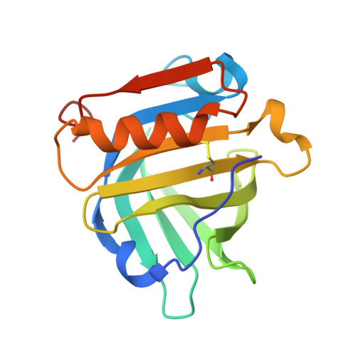
    3KZA

  • PubMed Abstract: 

    β-lactoglobulin (LG) contains nine β-strands (strands A-I) and one α-helix. Strands A-H form a β-barrel. At neutral pH, equine LG (ELG) is monomeric, whereas bovine LG (BLG) is dimeric, and the I-strands of its two subunits form an intermolecular β-sheet. We previously constructed a chimeric ELG in which the sequence of the I-strand was replaced with that of BLG. This chimera did not dimerize. For this study, we constructed the new chimera we call Gyuba (which means cow and horse in Japanese). The amino acid sequence of Gyuba includes the sequences of the BLG secondary structures and those of the ELG loops. The crystal structure of Gyuba is very similar to that of BLG and indicates that Gyuba dimerizes via the intermolecular β-sheet formed by the two I-strands. Thus, the entire arrangement of the secondary structural elements is important for LG dimer formation.


  • Organizational Affiliation

    Department of Bioinformatics, Soka University, 1-236 Tangi-cho, Hachioji, Tokyo 192-8577, Japan.


Macromolecules
Find similar proteins by:  (by identity cutoff)  |  3D Structure
Entity ID: 1
MoleculeChains Sequence LengthOrganismDetailsImage
Beta-lactoglobulin
A, B
162Equus caballusBos taurusMutation(s): 2 
Gene Names: LGB1
UniProt
Find proteins for P02758 (Equus caballus)
Explore P02758 
Go to UniProtKB:  P02758
Find proteins for P02754 (Bos taurus)
Explore P02754 
Go to UniProtKB:  P02754
Entity Groups  
Sequence Clusters30% Identity50% Identity70% Identity90% Identity95% Identity100% Identity
UniProt GroupsP02758P02754
Sequence Annotations
Expand
  • Reference Sequence
Experimental Data & Validation

Experimental Data

  • Method: X-RAY DIFFRACTION
  • Resolution: 2.00 Å
  • R-Value Free: 0.285 
  • R-Value Work: 0.235 
  • R-Value Observed: 0.237 
  • Space Group: P 65
Unit Cell:
Length ( Å )Angle ( ˚ )
a = 68.925α = 90
b = 68.925β = 90
c = 151.616γ = 120
Software Package:
Software NamePurpose
HKL-2000data collection
MOLREPphasing
REFMACrefinement
HKL-2000data reduction
HKL-2000data scaling

Structure Validation

View Full Validation Report


Entry History 

Deposition Data

Revision History  (Full details and data files)

  • Version 1.0: 2010-12-22
    Type: Initial release
  • Version 1.1: 2011-07-13
    Changes: Version format compliance
  • Version 1.2: 2011-11-09
    Changes: Database references
  • Version 1.3: 2017-08-23
    Changes: Refinement description, Source and taxonomy
  • Version 1.4: 2021-11-10
    Changes: Database references