


Crystal structure of the Tudor domains from Fragile X mental retardation syndrome-related protein 1
Guo, Y.H., Adams-Cioaba, M., Bian, C.B., Min, J.R., Structural Genomics Consortium (SGC)To be published.
Experimental Data Snapshot
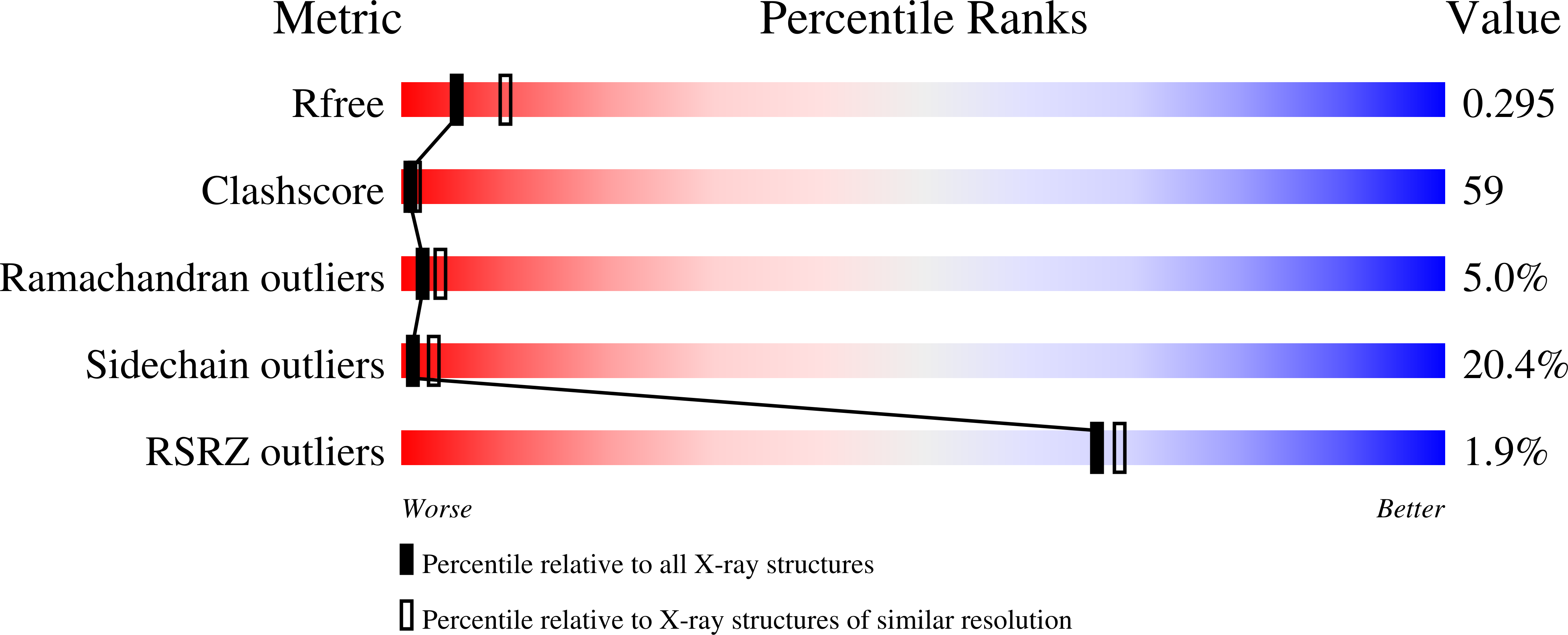
wwPDB Validation 3D Report Full Report
Entity ID: 1 | |||||
|---|---|---|---|---|---|
| Molecule | Chains | Sequence Length | Organism | Details | Image |
| Fragile X mental retardation syndrome-related protein 1 | 131 | Homo sapiens | Mutation(s): 0 Gene Names: FXR1 | 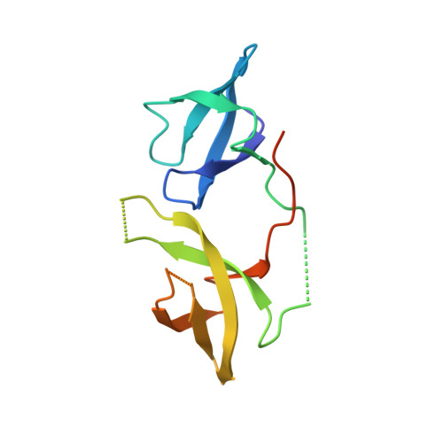 | |
UniProt & NIH Common Fund Data Resources | |||||
Find proteins for P51114 (Homo sapiens) Explore P51114 Go to UniProtKB: P51114 | |||||
PHAROS: P51114 GTEx: ENSG00000114416 | |||||
Entity Groups | |||||
| Sequence Clusters | 30% Identity50% Identity70% Identity90% Identity95% Identity100% Identity | ||||
| UniProt Group | P51114 | ||||
Sequence AnnotationsExpand | |||||
| |||||
| Ligands 2 Unique | |||||
|---|---|---|---|---|---|
| ID | Chains | Name / Formula / InChI Key | 2D Diagram | 3D Interactions | |
| DTT Query on DTT | B [auth A] | 2,3-DIHYDROXY-1,4-DITHIOBUTANE C4 H10 O2 S2 VHJLVAABSRFDPM-IMJSIDKUSA-N |  | ||
| GOL Query on GOL | C [auth A], D [auth A], E [auth A], F [auth A], G [auth A] | GLYCEROL C3 H8 O3 PEDCQBHIVMGVHV-UHFFFAOYSA-N |  | ||
| Length ( Å ) | Angle ( ˚ ) |
|---|---|
| a = 73.102 | α = 90 |
| b = 73.102 | β = 90 |
| c = 94.412 | γ = 120 |
| Software Name | Purpose |
|---|---|
| REFMAC | refinement |
RCSB PDB (citation) is hosted by
RCSB PDB is a member of the

