3KJ7

Crystal Structure of the Complex of C-lobe of Bovine Lactoferrin with Dextrin at 1.9 A Resolution


Experimental Data Snapshot

  • Method: X-RAY DIFFRACTION
  • Resolution: 1.91 Å
  • R-Value Free: 0.241 
  • R-Value Work: 0.209 
  • R-Value Observed: 0.211 

wwPDB Validation   3D Report Full Report


This is version 2.1 of the entry. See complete history


Literature

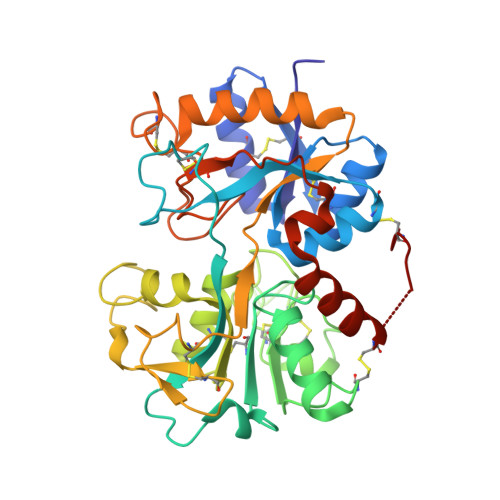
Specific interactions of C-terminal half (C-lobe) of lactoferrin protein with edible sugars: binding and structural studies with implications on diabetes.

Mir, R.Kumar, R.P.Singh, N.Vikram, G.P.Sinha, M.Bhushan, A.Kaur, P.Srinivasan, A.Sharma, S.Singh, T.P.

(2010) Int J Biol Macromol 47: 50-59

  • DOI: https://doi.org/10.1016/j.ijbiomac.2010.03.021
  • Primary Citation of Related Structures:  
    3K0V, 3KJ7

  • PubMed Abstract: 

    Bovine lactoferrin has been shown to reduce the levels of glucose in both normal subjects and non-insulin dependent diabetic patients. The binding studies have shown that various sugar molecules interact with lactoferrin indicating the presence of a sugar-binding site in the protein. Structural studies have revealed that the sugar-binding site is located in the C-terminal half (C-lobe) of bilobal lactoferrin. Since the sugar-binding site was part of the C-lobe, it was better to carry out binding and structural studies using C-lobe rather than the full protein molecule. Therefore, C-lobe was prepared by limited proteolysis of lactoferrin with enzyme proteinase K. It was purified to homogeneity for further studies. The addition of C-lobe to human serum showed significant lowering of glucose levels. The binding studies using C-lobe with nine sugars, glucose, galactose, mannose, xylose, maltose, cellobiose, lactose, sucrose and dextrin gave values of binding constants in the range of 10(-4) to 10(-5)M. The structure determinations of the complexes of C-lobe with all the nine sugars showed that all of them interact with C-lobe through the same recognition site involving several hydrogen bonds and van der Waals interactions.


  • Organizational Affiliation

    Department of Biophysics, All India Institute of Medical Sciences, Ansari Nagar, New Delhi 110029, India.


Macromolecules
Find similar proteins by:  (by identity cutoff)  |  3D Structure
Entity ID: 1
MoleculeChains Sequence LengthOrganismDetailsImage
Lactotransferrin345Bos taurusMutation(s): 0 
EC: 3.4.21
UniProt
Find proteins for P24627 (Bos taurus)
Explore P24627 
Go to UniProtKB:  P24627
Entity Groups  
Sequence Clusters30% Identity50% Identity70% Identity90% Identity95% Identity100% Identity
UniProt GroupP24627
Sequence Annotations
Expand
  • Reference Sequence
Oligosaccharides

Help

Entity ID: 2
MoleculeChains Length2D Diagram Glycosylation3D Interactions
2-acetamido-2-deoxy-beta-D-glucopyranose-(1-4)-2-acetamido-2-deoxy-beta-D-glucopyranose
B
2N-Glycosylation
Glycosylation Resources
GlyTouCan:  G42666HT
GlyCosmos:  G42666HT
GlyGen:  G42666HT
Entity ID: 3
MoleculeChains Length2D Diagram Glycosylation3D Interactions
alpha-D-mannopyranose-(1-4)-2-acetamido-2-deoxy-beta-D-glucopyranose-(1-4)-2-acetamido-2-deoxy-beta-D-glucopyranose
C, D
3N-Glycosylation
Glycosylation Resources
GlyTouCan:  G62182OO
GlyCosmos:  G62182OO
GlyGen:  G62182OO
Entity ID: 4
MoleculeChains Length2D Diagram Glycosylation3D Interactions
beta-D-glucopyranose-(1-4)-beta-D-glucopyranose-(1-4)-alpha-D-glucopyranose
E
3N/A
Glycosylation Resources
GlyTouCan:  G03861VK
GlyCosmos:  G03861VK
GlyGen:  G03861VK
Biologically Interesting Molecules (External Reference) 1 Unique
Experimental Data & Validation

Experimental Data

  • Method: X-RAY DIFFRACTION
  • Resolution: 1.91 Å
  • R-Value Free: 0.241 
  • R-Value Work: 0.209 
  • R-Value Observed: 0.211 
  • Space Group: P 1 21 1
Unit Cell:
Length ( Å )Angle ( ˚ )
a = 61.81α = 90
b = 50.134β = 107.1
c = 65.545γ = 90
Software Package:
Software NamePurpose
DENZOdata reduction
MOLREPphasing
REFMACrefinement
AUTOMARdata reduction
SCALEPACKdata scaling

Structure Validation

View Full Validation Report


Entry History 

Deposition Data

Revision History  (Full details and data files)

  • Version 1.0: 2010-02-16
    Type: Initial release
  • Version 1.1: 2011-07-13
    Changes: Version format compliance
  • Version 1.2: 2011-09-07
    Changes: Database references
  • Version 2.0: 2020-07-29
    Type: Remediation
    Reason: Carbohydrate remediation
    Changes: Atomic model, Data collection, Derived calculations, Non-polymer description, Structure summary
  • Version 2.1: 2023-09-06
    Changes: Data collection, Database references, Refinement description, Structure summary