3IOM

Crystal structure of Purine Nucleoside Phosphorylase from Mycobacterium tuberculosis in complex with 2'-Deoxyguanosine


Experimental Data Snapshot

  • Method: X-RAY DIFFRACTION
  • Resolution: 2.14 Å
  • R-Value Free: 0.249 
  • R-Value Work: 0.175 
  • R-Value Observed: 0.179 

wwPDB Validation   3D Report Full Report


Ligand Structure Quality Assessment 


This is version 1.2 of the entry. See complete history


Literature

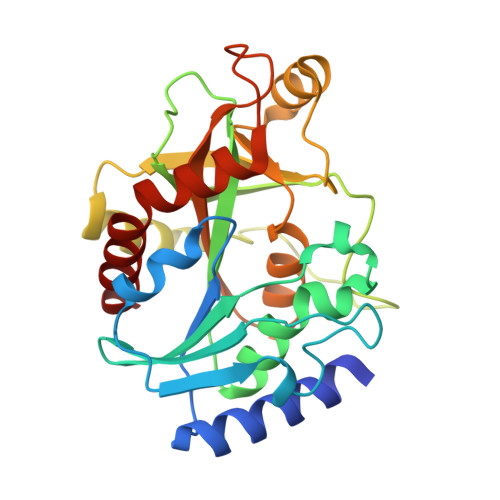
Crystallographic and docking studies of purine nucleoside phosphorylase from Mycobacterium tuberculosis.

Ducati, R.G.Basso, L.A.Santos, D.S.de Azevedo, W.F.

(2010) Bioorg Med Chem 18: 4769-4774

  • DOI: https://doi.org/10.1016/j.bmc.2010.05.009
  • Primary Citation of Related Structures:  
    3IOM

  • PubMed Abstract: 

    This work describes for the first time the structure of purine nucleoside phosphorylase from Mycobacterium tuberculosis (MtPNP) in complex with sulfate and its natural substrate, 2'-deoxyguanosine, and its application to virtual screening. We report docking studies of a set of molecules against this structure. Application of polynomial empirical scoring function was able to rank docking solutions with good predicting power which opens the possibility to apply this new criterion to analyze docking solutions and screen small-molecule databases for new chemical entities to inhibit MtPNP.


  • Organizational Affiliation

    Centro de Pesquisas em Biologia Molecular e Funcional, Instituto de Pesquisas Biomédicas, Pontifícia Universidade Católica do Rio Grande do Sul (PUCRS), Avenida Ipiranga 6681/92-A, 90619-900 Porto Alegre, RS, Brazil.


Macromolecules
Find similar proteins by:  (by identity cutoff)  |  3D Structure
Entity ID: 1
MoleculeChains Sequence LengthOrganismDetailsImage
Purine nucleoside phosphorylase
A, B
268Mycobacterium tuberculosisMutation(s): 0 
Gene Names: deoDMT3406MTV016.06punApunA (deoD) (Rv3307) (MT3406) (MTV016.06)Rv3307
EC: 2.4.2.1
UniProt
Find proteins for P9WP01 (Mycobacterium tuberculosis (strain ATCC 25618 / H37Rv))
Explore P9WP01 
Go to UniProtKB:  P9WP01
Entity Groups  
Sequence Clusters30% Identity50% Identity70% Identity90% Identity95% Identity100% Identity
UniProt GroupP9WP01
Sequence Annotations
Expand
  • Reference Sequence
Experimental Data & Validation

Experimental Data

  • Method: X-RAY DIFFRACTION
  • Resolution: 2.14 Å
  • R-Value Free: 0.249 
  • R-Value Work: 0.175 
  • R-Value Observed: 0.179 
  • Space Group: H 3
Unit Cell:
Length ( Å )Angle ( ˚ )
a = 113.48α = 90
b = 113.48β = 90
c = 85.011γ = 120
Software Package:
Software NamePurpose
MAR345dtbdata collection
AMoREphasing
REFMACrefinement
MOSFLMdata reduction
SCALAdata scaling

Structure Validation

View Full Validation Report


Ligand Structure Quality Assessment 


Entry History 

Deposition Data

Revision History  (Full details and data files)

  • Version 1.0: 2011-01-12
    Type: Initial release
  • Version 1.1: 2011-07-13
    Changes: Version format compliance
  • Version 1.2: 2023-09-06
    Changes: Data collection, Database references, Derived calculations, Refinement description