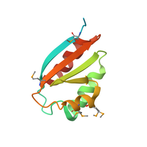
Crystal Structure of a Phosphocarrier Protein HPr from Bacillus anthracis str. Ames
Brunzelle, J.S., Anderson, S.M., Wawrzak, Z., Skarina, T., Onopriyenko, O., Savchenko, A., Anderson, W.F., Center for Structural Genomics of Infectious Diseases (CSGID)To be published.