3GWN

Crystal structure of the FAD binding domain from mimivirus sulfhydryl oxidase R596


Experimental Data Snapshot

  • Method: X-RAY DIFFRACTION
  • Resolution: 1.78 Å
  • R-Value Free: 0.213 
  • R-Value Work: 0.181 

wwPDB Validation   3D Report Full Report


Ligand Structure Quality Assessment 


This is version 1.3 of the entry. See complete history


Literature

Dimer interface migration in a viral sulfhydryl oxidase

Hakim, M.Fass, D.

(2009) J Mol Biol 391: 758-768

  • DOI: https://doi.org/10.1016/j.jmb.2009.06.070
  • Primary Citation of Related Structures:  
    3GWL, 3GWN

  • PubMed Abstract: 

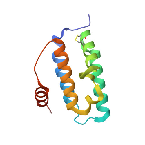
    Large double-stranded DNA viruses, including poxviruses and mimiviruses, encode enzymes to catalyze the formation of disulfide bonds in viral proteins produced in the cell cytosol, an atypical location for oxidative protein folding. These viral disulfide catalysts belong to a family of sulfhydryl oxidases that are dimers of a small five-helix fold containing a Cys-X-X-Cys motif juxtaposed to a flavin adenine dinucleotide cofactor. We report that the sulfhydryl oxidase pB119L from African swine fever virus (ASFV) uses for self-assembly surface different from that observed in homologs from mammals, plants, and fungi. Within a protein family, different packing interfaces for the same oligomerization state are extremely rare. We find that the alternate dimerization mode seen in ASFV pB119L is not characteristic of all viral sulfhydryl oxidases, as the flavin-binding domain from a mimivirus sulfhydryl oxidase assumes the same dimer structure as the known eukaryotic enzymes. ASFV pB119L demonstrates the potential of large double-stranded DNA viruses, which have faster mutation rates than their hosts and the tendency to incorporate host genes, to pioneer new protein folds and self-assembly modes.


  • Organizational Affiliation

    Department of Structural Biology, Weizmann Institute of Science, Rehovot, Israel.


Macromolecules
Find similar proteins by:  (by identity cutoff)  |  3D Structure
Entity ID: 1
MoleculeChains Sequence LengthOrganismDetailsImage
Probable FAD-linked sulfhydryl oxidase R596
A, B
114Acanthamoeba polyphaga mimivirusMutation(s): 0 
Gene Names: MIMI_R596R596
EC: 1.8.3.2
UniProt
Find proteins for Q5UP54 (Acanthamoeba polyphaga mimivirus)
Explore Q5UP54 
Go to UniProtKB:  Q5UP54
Entity Groups  
Sequence Clusters30% Identity50% Identity70% Identity90% Identity95% Identity100% Identity
UniProt GroupQ5UP54
Sequence Annotations
Expand
  • Reference Sequence
Experimental Data & Validation

Experimental Data

  • Method: X-RAY DIFFRACTION
  • Resolution: 1.78 Å
  • R-Value Free: 0.213 
  • R-Value Work: 0.181 
  • Space Group: P 21 21 21
Unit Cell:
Length ( Å )Angle ( ˚ )
a = 35.166α = 90
b = 58.584β = 90
c = 119.085γ = 90
Software Package:
Software NamePurpose
PHASERphasing
CNSrefinement
HKL-2000data reduction
HKL-2000data scaling

Structure Validation

View Full Validation Report


Ligand Structure Quality Assessment 


Entry History 

Deposition Data

Revision History  (Full details and data files)

  • Version 1.0: 2009-07-07
    Type: Initial release
  • Version 1.1: 2011-07-13
    Changes: Non-polymer description, Version format compliance
  • Version 1.2: 2016-12-07
    Changes: Database references
  • Version 1.3: 2023-11-01
    Changes: Data collection, Database references, Derived calculations, Refinement description