3GFI

Crystal structure of ST1710 complexed with its promoter DNA


Experimental Data Snapshot

  • Method: X-RAY DIFFRACTION
  • Resolution: 2.10 Å
  • R-Value Free: 0.280 
  • R-Value Work: 0.234 
  • R-Value Observed: 0.234 

wwPDB Validation   3D Report Full Report


This is version 1.3 of the entry. See complete history


Literature

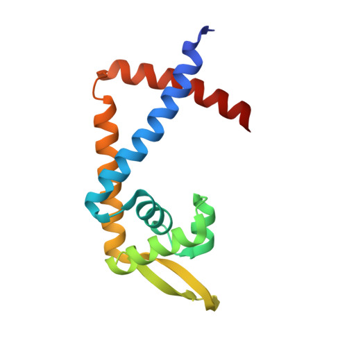
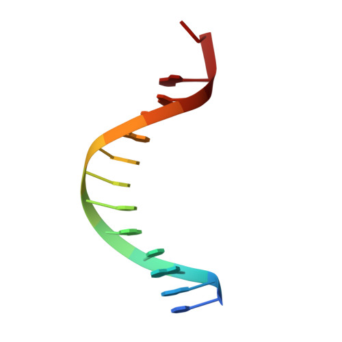
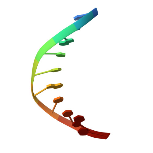
ST1710-DNA complex crystal structure reveals the DNA binding mechanism of the MarR family of regulators.

Kumarevel, T.Tanaka, T.Umehara, T.Yokoyama, S.

(2009) Nucleic Acids Res 37: 4723-4735

  • DOI: https://doi.org/10.1093/nar/gkp496
  • Primary Citation of Related Structures:  
    3GEZ, 3GF2, 3GFI, 3GFJ, 3GFL, 3GFM

  • PubMed Abstract: 

    ST1710, a member of the multiple antibiotic resistance regulator (MarR) family of regulatory proteins in bacteria and archaea, plays important roles in development of antibiotic resistance, a global health problem. Here, we present the crystal structure of ST1710 from Sulfolobus tokodaii strain 7 complexed with salicylate, a well-known inhibitor of MarR proteins and the ST1710 complex with its promoter DNA, refined to 1.8 and 2.10 A resolutions, respectively. The ST1710-DNA complex shares the topology of apo-ST1710 and MarR proteins, with each subunit containing a winged helix-turn-helix (wHtH) DNA binding motif. Significantly large conformational changes occurred upon DNA binding and in each of the dimeric monomers in the asymmetric unit of the ST1710-DNA complex. Conserved wHtH loop residues interacting with the bound DNA and mutagenic analysis indicated that R89, R90 and K91 were important for DNA recognition. Significantly, the bound DNA exhibited a new binding mechanism.


  • Organizational Affiliation

    RIKEN SPring-8 Center, Harima Institute, 1-1-1 Kouto, Sayo, Hyogo 679-5148, Japan. tskvel@spring8.or.jp


Macromolecules

Find similar proteins by:  (by identity cutoff)  |  3D Structure
Entity ID: 1
MoleculeChains Sequence LengthOrganismDetailsImage
146aa long hypothetical transcriptional regulatorA,
B [auth C]
146Sulfurisphaera tokodaiiMutation(s): 0 
Gene Names: ST1710
UniProt
Find proteins for Q96ZY1 (Sulfurisphaera tokodaii (strain DSM 16993 / JCM 10545 / NBRC 100140 / 7))
Explore Q96ZY1 
Go to UniProtKB:  Q96ZY1
Entity Groups  
Sequence Clusters30% Identity50% Identity70% Identity90% Identity95% Identity100% Identity
UniProt GroupQ96ZY1
Sequence Annotations
Expand
  • Reference Sequence

Find similar nucleic acids by:  Sequence   |   3D Structure  

Entity ID: 2
MoleculeChains LengthOrganismImage
5'-D(*TP*AP*AP*CP*AP*AP*TP*AP*GP*CP*AP*AP*A)-3'C [auth D]13N/A
Sequence Annotations
Expand
  • Reference Sequence

Find similar nucleic acids by:  Sequence   |   3D Structure  

Entity ID: 3
MoleculeChains LengthOrganismImage
5'-D(*TP*TP*GP*CP*TP*AP*TP*TP*GP*T)-3'D [auth E]10N/A
Sequence Annotations
Expand
  • Reference Sequence
Experimental Data & Validation

Experimental Data

  • Method: X-RAY DIFFRACTION
  • Resolution: 2.10 Å
  • R-Value Free: 0.280 
  • R-Value Work: 0.234 
  • R-Value Observed: 0.234 
  • Space Group: C 2 2 21
Unit Cell:
Length ( Å )Angle ( ˚ )
a = 94.44α = 90
b = 106.73β = 90
c = 82.26γ = 90
Software Package:
Software NamePurpose
CNSrefinement
PDB_EXTRACTdata extraction
CrystalCleardata collection
HKL-2000data reduction
HKL-2000data scaling
SOLVEphasing

Structure Validation

View Full Validation Report


Entry History 

Deposition Data

Revision History  (Full details and data files)

  • Version 1.0: 2009-08-25
    Type: Initial release
  • Version 1.1: 2011-07-13
    Changes: Version format compliance
  • Version 1.2: 2013-10-09
    Changes: Derived calculations
  • Version 1.3: 2024-03-20
    Changes: Data collection, Database references