3GFB

L-Threonine Dehydrogenase (TkTDH) from the hyperthermophilic archaeon Thermococcus kodakaraensis


Experimental Data Snapshot

  • Method: X-RAY DIFFRACTION
  • Resolution: 2.40 Å
  • R-Value Free: 0.277 
  • R-Value Work: 0.217 
  • R-Value Observed: 0.220 

wwPDB Validation   3D Report Full Report


Ligand Structure Quality Assessment 


This is version 1.2 of the entry. See complete history


Literature

Structure and function of the l-threonine dehydrogenase (TkTDH) from the hyperthermophilic archaeon Thermococcus kodakaraensis

Bowyer, A.Mikolajek, H.Stuart, J.W.Wood, S.P.Jamil, F.Rashid, N.Akhtar, M.Cooper, J.B.

(2009) J Struct Biol 168: 294-304

  • DOI: https://doi.org/10.1016/j.jsb.2009.07.011
  • Primary Citation of Related Structures:  
    3GFB

  • PubMed Abstract: 

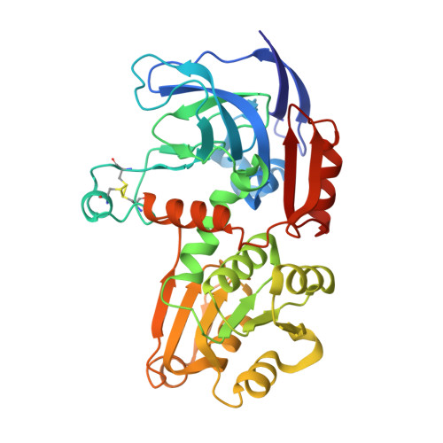
    The X-ray structure of the holo-form of l-threonine dehydrogenase (TDH) from Thermococcus kodakaraensis (TkTDH) has been determined at 2.4A resolution. TDH catalyses the NAD(+)-dependent oxidation of l-threonine to 2-amino-3-ketobutyrate, and is one of the first enzymes in this family to be solved by X-ray crystallography. The enzyme is a homo-tetramer, each monomer consisting of 350 amino acids that form two domains; a catalytic domain and a nicotinamide co-factor (NAD(+))-binding domain, which contains an alpha/beta Rossmann fold motif. An extended twelve-stranded beta-sheet is formed by the association of pairs of monomers in the tetramer. TkTDH shows strong overall structural similarity to TDHs from thermophiles and alcohol dehydrogenases (ADH) from lower life forms, despite low sequence homology, exhibiting the same overall fold of the monomer and assembly of the tetramer. The structure reveals the binding site of the essential co-factor NAD(+) which is present in all subunits. Docking studies suggest a mode of interaction of TDH with 2-amino-3-ketobutyrate CoA ligase, the subsequent enzyme in the pathway for conversion of threonine to glycine. TDH is known to form a stable functional complex with 2-amino-3-ketobutyrate ligase, most probably to shield an unstable intermediate.


  • Organizational Affiliation

    University of Southampton, UK.


Macromolecules
Find similar proteins by:  (by identity cutoff)  |  3D Structure
Entity ID: 1
MoleculeChains Sequence LengthOrganismDetailsImage
L-threonine 3-dehydrogenase
A, B, C, D
350Thermococcus kodakarensis KOD1Mutation(s): 0 
Gene Names: tdhTK0916
EC: 1.1.1.103
UniProt
Find proteins for Q5JI69 (Thermococcus kodakarensis (strain ATCC BAA-918 / JCM 12380 / KOD1))
Explore Q5JI69 
Go to UniProtKB:  Q5JI69
Entity Groups  
Sequence Clusters30% Identity50% Identity70% Identity90% Identity95% Identity100% Identity
UniProt GroupQ5JI69
Sequence Annotations
Expand
  • Reference Sequence
Small Molecules
Ligands 2 Unique
IDChains Name / Formula / InChI Key2D Diagram3D Interactions
NAD
Query on NAD

Download Ideal Coordinates CCD File 
E [auth A],
F [auth B],
L [auth C],
P [auth D]
NICOTINAMIDE-ADENINE-DINUCLEOTIDE
C21 H27 N7 O14 P2
BAWFJGJZGIEFAR-NNYOXOHSSA-N
SO4
Query on SO4

Download Ideal Coordinates CCD File 
G [auth B]
H [auth B]
I [auth B]
J [auth B]
K [auth B]
G [auth B],
H [auth B],
I [auth B],
J [auth B],
K [auth B],
M [auth C],
N [auth C],
O [auth C],
Q [auth D],
R [auth D]
SULFATE ION
O4 S
QAOWNCQODCNURD-UHFFFAOYSA-L
Experimental Data & Validation

Experimental Data

  • Method: X-RAY DIFFRACTION
  • Resolution: 2.40 Å
  • R-Value Free: 0.277 
  • R-Value Work: 0.217 
  • R-Value Observed: 0.220 
  • Space Group: P 43 21 2
Unit Cell:
Length ( Å )Angle ( ˚ )
a = 124.481α = 90
b = 124.481β = 90
c = 271.143γ = 90
Software Package:
Software NamePurpose
PHASERphasing
REFMACrefinement
MOSFLMdata reduction
SCALAdata scaling

Structure Validation

View Full Validation Report


Ligand Structure Quality Assessment 


Entry History 

Deposition Data

  • Released Date: 2009-10-06 
  • Deposition Author(s): Bowyer, A.

Revision History  (Full details and data files)

  • Version 1.0: 2009-10-06
    Type: Initial release
  • Version 1.1: 2011-07-13
    Changes: Version format compliance
  • Version 1.2: 2023-09-06
    Changes: Data collection, Database references, Derived calculations, Refinement description