3GA9

Crystal structure of Bacillus anthracis transpeptidase enzyme CapD, crystal form II


Experimental Data Snapshot

  • Method: X-RAY DIFFRACTION
  • Resolution: 2.30 Å
  • R-Value Free: 0.252 
  • R-Value Work: 0.197 
  • R-Value Observed: 0.200 

wwPDB Validation   3D Report Full Report


This is version 1.4 of the entry. See complete history


Literature

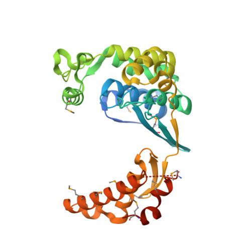
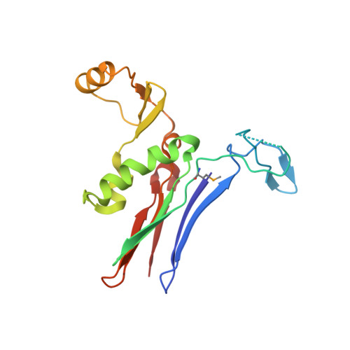
Crystal Structure of Bacillus anthracis Transpeptidase Enzyme CapD.

Wu, R.Richter, S.Zhang, R.G.Anderson, V.J.Missiakas, D.Joachimiak, A.

(2009) J Biol Chem 284: 24406-24414

  • DOI: https://doi.org/10.1074/jbc.M109.019034
  • Primary Citation of Related Structures:  
    3G9K, 3GA9

  • PubMed Abstract: 

    Bacillus anthracis elaborates a poly-gamma-d-glutamic acid capsule that protects bacilli from phagocytic killing during infection. The enzyme CapD generates amide bonds with peptidoglycan cross-bridges to anchor capsular material within the cell wall envelope of B. anthracis. The capsular biosynthetic pathway is essential for virulence during anthrax infections and can be targeted for anti-infective inhibition with small molecules. Here, we present the crystal structures of the gamma-glutamyltranspeptidase CapD with and without alpha-l-Glu-l-Glu dipeptide, a non-hydrolyzable analog of poly-gamma-d-glutamic acid, in the active site. Purified CapD displays transpeptidation activity in vitro, and its structure reveals an active site broadly accessible for poly-gamma-glutamate binding and processing. Using structural and biochemical information, we derive a mechanistic model for CapD catalysis whereby Pro(427), Gly(428), and Gly(429) activate the catalytic residue of the enzyme, Thr(352), and stabilize an oxyanion hole via main chain amide hydrogen bonds.


  • Organizational Affiliation

    Biosciences Division, Midwest Center for Structural Genomics and Structural Biology Center, Argonne National Laboratory, Argonne, Illinois 60439, USA.


Macromolecules
Find similar proteins by:  (by identity cutoff)  |  3D Structure
Entity ID: 1
MoleculeChains Sequence LengthOrganismDetailsImage
Capsule biosynthesis protein capDA [auth L]323Bacillus anthracisMutation(s): 0 
Gene Names: BAK_B0097BXB0063capDdepGBAA_pXO2_0063pXO2-55
UniProt
Find proteins for Q51693 (Bacillus anthracis)
Explore Q51693 
Go to UniProtKB:  Q51693
Entity Groups  
Sequence Clusters30% Identity50% Identity70% Identity90% Identity95% Identity100% Identity
UniProt GroupQ51693
Sequence Annotations
Expand
  • Reference Sequence
Find similar proteins by:  (by identity cutoff)  |  3D Structure
Entity ID: 2
MoleculeChains Sequence LengthOrganismDetailsImage
Capsule biosynthesis protein capDB [auth S]177Bacillus anthracisMutation(s): 0 
Gene Names: capDdeppXO2-55BXB0063GBAA_pXO2_0063
UniProt
Find proteins for Q51693 (Bacillus anthracis)
Explore Q51693 
Go to UniProtKB:  Q51693
Entity Groups  
Sequence Clusters30% Identity50% Identity70% Identity90% Identity95% Identity100% Identity
UniProt GroupQ51693
Sequence Annotations
Expand
  • Reference Sequence
Small Molecules
Modified Residues  1 Unique
IDChains TypeFormula2D DiagramParent
MSE
Query on MSE
A [auth L]L-PEPTIDE LINKINGC5 H11 N O2 SeMET
Experimental Data & Validation

Experimental Data

  • Method: X-RAY DIFFRACTION
  • Resolution: 2.30 Å
  • R-Value Free: 0.252 
  • R-Value Work: 0.197 
  • R-Value Observed: 0.200 
  • Space Group: P 21 21 21
Unit Cell:
Length ( Å )Angle ( ˚ )
a = 47.704α = 90
b = 72β = 90
c = 128.853γ = 90
Software Package:
Software NamePurpose
HKL-3000data collection
MLPHAREphasing
REFMACrefinement
HKL-3000data reduction
HKL-3000data scaling

Structure Validation

View Full Validation Report


Entry History 

Deposition Data

Revision History  (Full details and data files)

  • Version 1.0: 2009-06-16
    Type: Initial release
  • Version 1.1: 2011-07-13
    Changes: Advisory, Refinement description, Version format compliance
  • Version 1.2: 2011-10-19
    Changes: Structure summary
  • Version 1.3: 2011-11-02
    Changes: Structure summary
  • Version 1.4: 2018-01-24
    Changes: Structure summary