3GA5

X-ray structure of glucose/galactose receptor from Salmonella typhimurium in complex with (2R)-glyceryl-beta-D-galactopyranoside


Experimental Data Snapshot

  • Method: X-RAY DIFFRACTION
  • Resolution: 1.87 Å
  • R-Value Free: 0.222 
  • R-Value Work: 0.170 
  • R-Value Observed: 0.173 

wwPDB Validation   3D Report Full Report


Ligand Structure Quality Assessment 


This is version 1.3 of the entry. See complete history


Literature

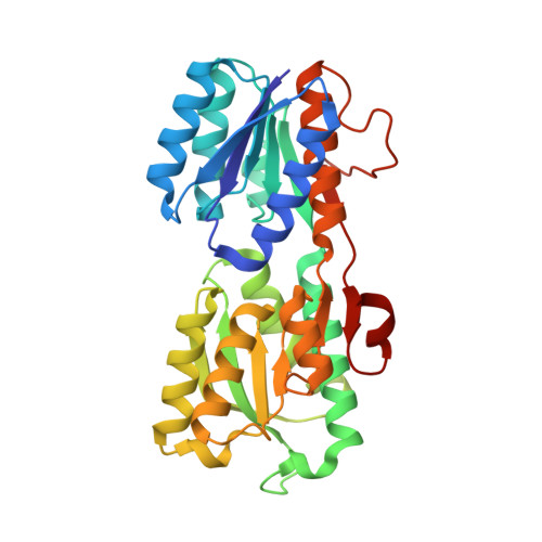
X-ray structure of glucose/galactose receptor from Salmonella typhimurium in complex with the physiological ligand, (2R)-glyceryl-beta-D-galactopyranoside

Sooriyaarachchi, S.Ubhayasekera, W.Boos, W.Mowbray, S.L.

(2009) FEBS J 276: 2116-2124

  • DOI: https://doi.org/10.1111/j.1742-4658.2009.06945.x
  • Primary Citation of Related Structures:  
    3GA5

  • PubMed Abstract: 

    Periplasmic binding proteins are abundant in bacteria by virtue of their essential roles as high-affinity receptors in ABC transport systems and chemotaxis. One of the best studied of these receptors is the so-called glucose/galactose-binding protein. Here, we report the X-ray structure of the Salmonella typhimurium protein bound to the physiologically relevant ligand, (2R)-glyceryl-beta-D-galactopyranoside, solved by molecular replacement, and refined to 1.87 A resolution with R and R-free values of 17% and 22%. The structure identifies three amino acid residues that are diagnostic of (2R)-glyceryl-beta-D-galactopyranoside binding (Thr110, Asp154 and Gln261), as opposed to binding to the monosaccharides glucose and galactose. These three residues are conserved in essentially all available glucose/galactose-binding protein sequences, indicating that the binding of (2R)-glyceryl-beta-D-galactopyranoside is the rule rather than the exception for receptors of this type. The role of (2R)-glyceryl-beta-D-galactopyranoside in bacterial biology is discussed. Further, comparison of the available structures provides the most complete description of the conformational changes of glucose/galactose-binding protein to date. The structures follow a smooth and continuous path from the most closed structure [that bound to (2R)-glyceryl-beta-D-galactopyranoside] to the most open (an apo structure).


  • Organizational Affiliation

    Department of Molecular Biology, Swedish University of Agricultural Sciences, Uppsala, Sweden.


Macromolecules
Find similar proteins by:  (by identity cutoff)  |  3D Structure
Entity ID: 1
MoleculeChains Sequence LengthOrganismDetailsImage
D-galactose-binding periplasmic protein
A, B
309Salmonella enterica subsp. enterica serovar TyphimuriumMutation(s): 0 
Gene Names: mglB
UniProt
Find proteins for P23905 (Salmonella typhimurium (strain LT2 / SGSC1412 / ATCC 700720))
Explore P23905 
Go to UniProtKB:  P23905
Entity Groups  
Sequence Clusters30% Identity50% Identity70% Identity90% Identity95% Identity100% Identity
UniProt GroupP23905
Sequence Annotations
Expand
  • Reference Sequence
Small Molecules
Ligands 4 Unique
IDChains Name / Formula / InChI Key2D Diagram3D Interactions
RGG
Query on RGG

Download Ideal Coordinates CCD File 
C [auth A],
G [auth B]
(2R)-2,3-dihydroxypropyl beta-D-galactopyranoside
C9 H18 O8
NHJUPBDCSOGIKX-NTXXKDEISA-N
SCN
Query on SCN

Download Ideal Coordinates CCD File 
E [auth A]THIOCYANATE ION
C N S
ZMZDMBWJUHKJPS-UHFFFAOYSA-M
CA
Query on CA

Download Ideal Coordinates CCD File 
D [auth A],
H [auth B]
CALCIUM ION
Ca
BHPQYMZQTOCNFJ-UHFFFAOYSA-N
NA
Query on NA

Download Ideal Coordinates CCD File 
F [auth A],
I [auth B]
SODIUM ION
Na
FKNQFGJONOIPTF-UHFFFAOYSA-N
Binding Affinity Annotations 
IDSourceBinding Affinity
RGG PDBBind:  3GA5 Kd: 3200 (nM) from 1 assay(s)
Binding MOAD:  3GA5 Kd: 3200 (nM) from 1 assay(s)
Experimental Data & Validation

Experimental Data

  • Method: X-RAY DIFFRACTION
  • Resolution: 1.87 Å
  • R-Value Free: 0.222 
  • R-Value Work: 0.170 
  • R-Value Observed: 0.173 
  • Space Group: P 21 21 21
Unit Cell:
Length ( Å )Angle ( ˚ )
a = 36.458α = 90
b = 109.275β = 90
c = 150.703γ = 90
Software Package:
Software NamePurpose
MOLREPphasing
REFMACrefinement
MOSFLMdata reduction
SCALAdata scaling

Structure Validation

View Full Validation Report


Ligand Structure Quality Assessment 


Entry History 

Deposition Data

Revision History  (Full details and data files)

  • Version 1.0: 2009-04-14
    Type: Initial release
  • Version 1.1: 2011-07-13
    Changes: Version format compliance
  • Version 1.2: 2020-07-29
    Type: Remediation
    Reason: Carbohydrate remediation
    Changes: Data collection, Derived calculations
  • Version 1.3: 2023-11-01
    Changes: Data collection, Database references, Refinement description, Structure summary