3FIL

Structural and energetic determinants for hyperstable variants of GB1 obtained from in-vitro evolution


Experimental Data Snapshot

  • Method: X-RAY DIFFRACTION
  • Resolution: 0.88 Å
  • R-Value Free: 0.149 
  • R-Value Work: 0.124 
  • R-Value Observed: 0.125 

wwPDB Validation   3D Report Full Report


This is version 1.4 of the entry. See complete history


Literature

Dimer Formation of a Stabilized Gbeta1 Variant: A Structural and Energetic Analysis

Thoms, S.Max, K.E.A.Wunderlich, M.Jacso, T.Lilie, H.Reif, B.Heinemann, U.Schmid, F.X.

(2009) J Mol Biol 391: 918-932

  • DOI: https://doi.org/10.1016/j.jmb.2009.06.031
  • Primary Citation of Related Structures:  
    3FIL

  • PubMed Abstract: 

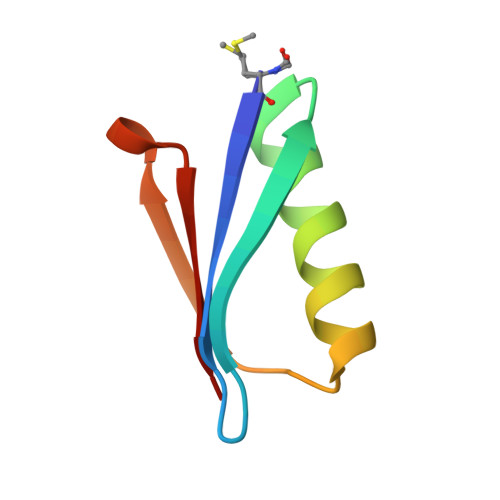
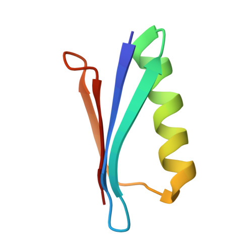
    In previous work, a strongly stabilized variant of the beta1 domain of streptococcal protein G (Gbeta1) was obtained by an in vitro selection method. This variant, termed Gbeta1-M2, contains the four substitutions E15V, T16L, T18I, and N37L. Here we elucidated the molecular basis of the observed strong stabilizations. The contributions of these four residues were analyzed individually and in various combinations, additional selections with focused Gbeta1 gene libraries were performed, and the crystal structure of Gbeta1-M2 was determined. All single substitutions (E15V, T16L, T18I, and N37L) stabilize wild-type Gbeta1 by contributions of between 1.6 and 6.0 kJ mol(-1) (at 70 degrees C). Hydrophobic residues at positions 16 and 37 provide the major contribution to stabilization by enlarging the hydrophobic core of Gbeta1. They also increase the tendency to form dimers, as shown by dependence on the concentration of apparent molecular mass in analytical ultracentrifugation, by concentration-dependent stability, and by a strongly increased van't Hoff enthalpy of unfolding. The 0.88-A crystal structure of Gbeta1-M2 and NMR measurements in solution provide the explanation for the observed dimer formation. It involves a head-to-head arrangement of two Gbeta1-M2 molecules via six intermolecular hydrogen bonds between the two beta strands 2 and 2' and an adjacent self-complementary hydrophobic surface area, which is created by the T16L and N37L substitutions and a large 120 degrees rotation of the Tyr33 side chain. This removal of hydrophilic groups and the malleability of the created hydrophobic surface provide the basis for the dimer formation of stabilized Gbeta1 variants.


  • Organizational Affiliation

    Universität Bayreuth, Germany.


Macromolecules
Find similar proteins by:  (by identity cutoff)  |  3D Structure
Entity ID: 1
MoleculeChains Sequence LengthOrganismDetailsImage
Immunoglobulin G-binding protein G56Streptococcus sp. 'group GMutation(s): 5 
Gene Names: spg
UniProt
Find proteins for P19909 (Streptococcus sp. group G)
Explore P19909 
Go to UniProtKB:  P19909
Entity Groups  
Sequence Clusters30% Identity50% Identity70% Identity90% Identity95% Identity100% Identity
UniProt GroupP19909
Sequence Annotations
Expand
  • Reference Sequence
Find similar proteins by:  (by identity cutoff)  |  3D Structure
Entity ID: 2
MoleculeChains Sequence LengthOrganismDetailsImage
Immunoglobulin G-binding protein G56Streptococcus sp. 'group GMutation(s): 5 
Gene Names: spg
UniProt
Find proteins for P19909 (Streptococcus sp. group G)
Explore P19909 
Go to UniProtKB:  P19909
Entity Groups  
Sequence Clusters30% Identity50% Identity70% Identity90% Identity95% Identity100% Identity
UniProt GroupP19909
Sequence Annotations
Expand
  • Reference Sequence
Small Molecules
Ligands 1 Unique
IDChains Name / Formula / InChI Key2D Diagram3D Interactions
CA
Query on CA

Download Ideal Coordinates CCD File 
C [auth A]CALCIUM ION
Ca
BHPQYMZQTOCNFJ-UHFFFAOYSA-N
Modified Residues  1 Unique
IDChains TypeFormula2D DiagramParent
FME
Query on FME
A
L-PEPTIDE LINKINGC6 H11 N O3 SMET
Experimental Data & Validation

Experimental Data

  • Method: X-RAY DIFFRACTION
  • Resolution: 0.88 Å
  • R-Value Free: 0.149 
  • R-Value Work: 0.124 
  • R-Value Observed: 0.125 
  • Space Group: P 21 21 21
Unit Cell:
Length ( Å )Angle ( ˚ )
a = 29.694α = 90
b = 38.93β = 90
c = 83.326γ = 90
Software Package:
Software NamePurpose
MAR345data collection
AMoREphasing
SHELXL-97refinement
XDSdata reduction
XSCALEdata scaling

Structure Validation

View Full Validation Report


Entry History 

Deposition Data

Revision History  (Full details and data files)

  • Version 1.0: 2009-08-18
    Type: Initial release
  • Version 1.1: 2011-07-13
    Changes: Version format compliance
  • Version 1.2: 2017-11-01
    Changes: Advisory, Refinement description
  • Version 1.3: 2021-11-10
    Changes: Advisory, Database references, Derived calculations
  • Version 1.4: 2023-11-01
    Changes: Data collection, Refinement description