3F8N

Crystal structure of PerR-Zn-Mn


Experimental Data Snapshot

  • Method: X-RAY DIFFRACTION
  • Resolution: 3.15 Å
  • R-Value Free: 0.315 
  • R-Value Work: 0.275 
  • R-Value Observed: 0.277 

wwPDB Validation   3D Report Full Report


This is version 1.2 of the entry. See complete history


Literature

Structural characterization of the active form of PerR: insights into the metal-induced activation of PerR and Fur proteins for DNA binding

Jacquamet, L.Traore, D.A.K.Ferrer, J.-L.Proux, O.Testemale, D.Hazemann, J.-L.Nazarenko, E.El Ghazouani, A.Caux-Thang, C.Duarte, V.Latour, J.-M.

(2009) Mol Microbiol 73: 20-31

  • DOI: https://doi.org/10.1111/j.1365-2958.2009.06753.x
  • Primary Citation of Related Structures:  
    3F8N

  • PubMed Abstract: 

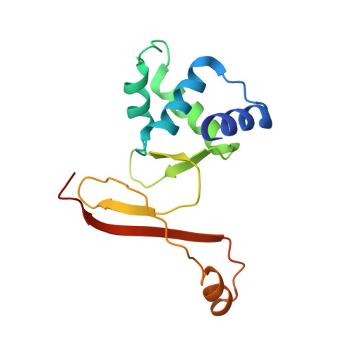
    In Bacillus subtilis, the transcription factor PerR is an iron dependant sensor of H(2)O(2). The sensing mechanism relies on a selective metal catalysed oxidation of two histidine residues of the regulatory site. Here we present the first crystal structure of the active PerR protein in complex with a Mn(2+) ion. In addition, X-ray absorption spectroscopy experiments were performed to characterize the corresponding iron form of the protein. Both studies reveal a penta-coordinate arrangement of the regulatory site that involves three histidines and two aspartates. One of the histidine ligand belongs to the N-terminal domain. Binding of this residue to the regulatory metal allows the protein to adopt a caliper-like conformation suited to DNA binding. Since this histidine is conserved in all PerR and a vast majority of Fur proteins, it is likely that the allosteric switch induced by the regulatory metal is general for this family of metalloregulators.


  • Organizational Affiliation

    Institut de Biologie Structurale CEA-CNRS-UJF, LCCP, GSY, 41 rue Jules Horowitz, 38027 Grenoble Cedex 1, France.


Macromolecules
Find similar proteins by:  (by identity cutoff)  |  3D Structure
Entity ID: 1
MoleculeChains Sequence LengthOrganismDetailsImage
Peroxide operon regulator
A, B
145Bacillus subtilisMutation(s): 0 
Gene Names: perR
UniProt
Find proteins for P71086 (Bacillus subtilis (strain 168))
Explore P71086 
Go to UniProtKB:  P71086
Entity Groups  
Sequence Clusters30% Identity50% Identity70% Identity90% Identity95% Identity100% Identity
UniProt GroupP71086
Sequence Annotations
Expand
  • Reference Sequence
Experimental Data & Validation

Experimental Data

  • Method: X-RAY DIFFRACTION
  • Resolution: 3.15 Å
  • R-Value Free: 0.315 
  • R-Value Work: 0.275 
  • R-Value Observed: 0.277 
  • Space Group: P 1 21 1
Unit Cell:
Length ( Å )Angle ( ˚ )
a = 49.3α = 90
b = 63.34β = 80.41
c = 66.73γ = 90
Software Package:
Software NamePurpose
ADSCdata collection
PHASERphasing
REFMACrefinement
XDSdata reduction
XSCALEdata scaling

Structure Validation

View Full Validation Report


Entry History 

Deposition Data

Revision History  (Full details and data files)

  • Version 1.0: 2009-06-16
    Type: Initial release
  • Version 1.1: 2011-07-13
    Changes: Version format compliance
  • Version 1.2: 2023-11-01
    Changes: Data collection, Database references, Derived calculations, Refinement description