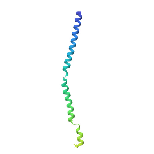
3F6N

Crystal structure of the virion-associated protein P3 from Caulimovirus


Experimental Data Snapshot

  • Method: X-RAY DIFFRACTION
  • Resolution: 3.10 Å
  • R-Value Free: 0.287 
  • R-Value Work: 0.222 
  • R-Value Observed: 0.229 

wwPDB Validation   3D Report Full Report


This is version 1.2 of the entry. See complete history


Literature

Structural insights into the molecular mechanisms of cauliflower mosaic virus transmission by its insect vector.

Hoh, F.Uzest, M.Drucker, M.Plisson-Chastang, C.Bron, P.Blanc, S.Dumas, C.

(2010) J Virol 84: 4706-4713

  • DOI: https://doi.org/10.1128/JVI.02662-09
  • Primary Citation of Related Structures:  
    3F6N, 3K4T

  • PubMed Abstract: 

    Cauliflower mosaic virus (CaMV) is transmitted from plant to plant through a seemingly simple interaction with insect vectors. This process involves an aphid receptor and two viral proteins, P2 and P3. P2 binds to both the aphid receptor and P3, itself tightly associated with the virus particle, with the ensemble forming a transmissible viral complex. Here, we describe the conformations of both unliganded CaMV P3 protein and its virion-associated form. X-ray crystallography revealed that the N-terminal domain of unliganded P3 is a tetrameric parallel coiled coil with a unique organization showing two successive four-stranded subdomains with opposite supercoiling handedness stabilized by a ring of interchain disulfide bridges. A structural model of virus-liganded P3 proteins, folding as an antiparallel coiled-coil network coating the virus surface, was derived from molecular modeling. Our results highlight the structural and biological versatility of this coiled-coil structure and provide new insights into the molecular mechanisms involved in CaMV acquisition and transmission by the insect vector.


  • Organizational Affiliation

    Centre de Biochimie Structurale, 29 rue de Navacelles, 34090 Montpellier Cedex, France.


Macromolecules
Find similar proteins by:  (by identity cutoff)  |  3D Structure
Entity ID: 1
MoleculeChains Sequence LengthOrganismDetailsImage
Virion-associated protein
A, B, C, D
129Cauliflower mosaic virus (STRAIN STRASBOURG)Mutation(s): 0 
Gene Names: ORF IIIORF-3
UniProt
Find proteins for P03551 (Cauliflower mosaic virus (strain Strasbourg))
Explore P03551 
Go to UniProtKB:  P03551
Entity Groups  
Sequence Clusters30% Identity50% Identity70% Identity90% Identity95% Identity100% Identity
UniProt GroupP03551
Sequence Annotations
Expand
  • Reference Sequence
Experimental Data & Validation

Experimental Data

  • Method: X-RAY DIFFRACTION
  • Resolution: 3.10 Å
  • R-Value Free: 0.287 
  • R-Value Work: 0.222 
  • R-Value Observed: 0.229 
  • Space Group: P 64
Unit Cell:
Length ( Å )Angle ( ˚ )
a = 104.943α = 90
b = 104.943β = 90
c = 72.527γ = 120
Software Package:
Software NamePurpose
MOSFLMdata reduction
SCALAdata scaling
PHASERphasing
REFMACrefinement
PDB_EXTRACTdata extraction
ADSCdata collection

Structure Validation

View Full Validation Report


Entry History 

Deposition Data

Revision History  (Full details and data files)

  • Version 1.0: 2009-11-24
    Type: Initial release
  • Version 1.1: 2011-07-13
    Changes: Advisory, Refinement description, Version format compliance
  • Version 1.2: 2023-11-01
    Changes: Data collection, Database references, Refinement description