3EQ7

Prolyl oligopeptidase complexed with R-Pro-(decarboxy-Pro)-Type inhibitors


Experimental Data Snapshot

  • Method: X-RAY DIFFRACTION
  • Resolution: 2.89 Å
  • R-Value Free: 0.257 
  • R-Value Work: 0.178 

wwPDB Validation   3D Report Full Report


Ligand Structure Quality Assessment 


This is version 1.4 of the entry. See complete history


Literature

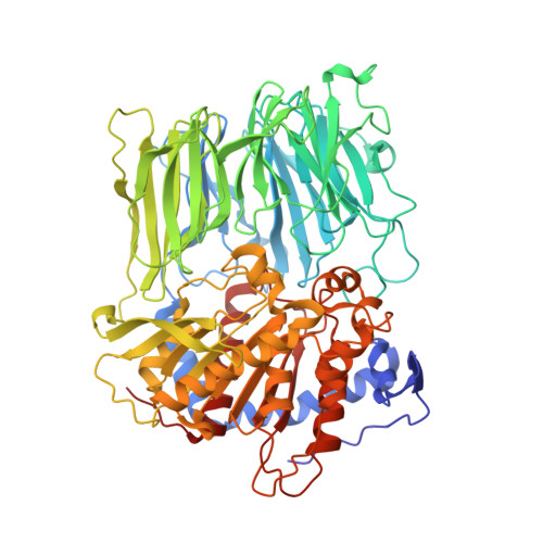
Prolyl oligopeptidase inhibition by N-acyl-pro-pyrrolidine-type molecules

Kanai, K.Aranyi, P.Bocskei, Z.Ferenczy, G.Harmat, V.Simon, K.Batori, S.Naray-Szabo, G.Hermecz, I.

(2008) J Med Chem 51: 7514-7522

  • DOI: https://doi.org/10.1021/jm800944x
  • Primary Citation of Related Structures:  
    3EQ7, 3EQ8, 3EQ9

  • PubMed Abstract: 

    Three novel, N-acyl-pro-pyrrolidine-type, inhibitors of prolyl oligopeptidase (POP) with nanomolar activities were synthesized and their binding analyzed to the host enzyme in the light of X-ray diffraction and molecular modeling studies. We were interested in the alteration in the binding affinity at the S3 site as a function of the properties of the N-terminal group of the inhibitors. Our studies revealed that, for inhibitors with flat aromatic terminal groups, the optimal length of the linker chain is three C-C bonds, but this increases to four C-C bonds if there is a bulky group in the terminal position. Molecular dynamics calculations indicate that this is due to the better fit into the binding pocket. A 4-fold enhancement of the inhibitor activity upon replacement of the 4-CH2 group of the proline ring by CF2 is a consequence of a weak hydrogen bond formed between the fluorine atom and the hydroxy group of Tyr473 of the host enzyme. There is notably good agreement between the calculated and experimental free energies of binding; the average error in the IC50 values is around 1 order of magnitude.


  • Organizational Affiliation

    CHINOIN, Ltd, H-1045 Budapest, Hungary.


Macromolecules
Find similar proteins by:  (by identity cutoff)  |  3D Structure
Entity ID: 1
MoleculeChains Sequence LengthOrganismDetailsImage
Prolyl endopeptidase710Sus scrofaMutation(s): 0 
EC: 3.4.21.26
UniProt
Find proteins for P23687 (Sus scrofa)
Explore P23687 
Go to UniProtKB:  P23687
Entity Groups  
Sequence Clusters30% Identity50% Identity70% Identity90% Identity95% Identity100% Identity
UniProt GroupP23687
Sequence Annotations
Expand
  • Reference Sequence
Small Molecules
Ligands 1 Unique
IDChains Name / Formula / InChI Key2D Diagram3D Interactions
X99
Query on X99

Download Ideal Coordinates CCD File 
B [auth A]2-{3-[(2S)-4,4-difluoro-2-(pyrrolidin-1-ylcarbonyl)pyrrolidin-1-yl]-3-oxopropyl}-isoindole-1,3(2H)-dione
C20 H21 F2 N3 O4
ZSXNPAWXICXNGZ-HNNXBMFYSA-N
Binding Affinity Annotations 
IDSourceBinding Affinity
X99 BindingDB:  3EQ7 IC50: 0.81 (nM) from 1 assay(s)
PDBBind:  3EQ7 IC50: 0.81 (nM) from 1 assay(s)
Experimental Data & Validation

Experimental Data

  • Method: X-RAY DIFFRACTION
  • Resolution: 2.89 Å
  • R-Value Free: 0.257 
  • R-Value Work: 0.178 
  • Space Group: P 21 21 21
Unit Cell:
Length ( Å )Angle ( ˚ )
a = 72.44α = 90
b = 101.44β = 90
c = 112.25γ = 90
Software Package:
Software NamePurpose
bioteXdata collection
AMoREphasing
X-PLORrefinement
bioteXdata reduction
SCALAdata scaling

Structure Validation

View Full Validation Report


Ligand Structure Quality Assessment 


Entry History 

Deposition Data

Revision History  (Full details and data files)

  • Version 1.0: 2009-08-25
    Type: Initial release
  • Version 1.1: 2011-07-13
    Changes: Version format compliance
  • Version 1.2: 2012-01-18
    Changes: Non-polymer description
  • Version 1.3: 2017-10-25
    Changes: Refinement description
  • Version 1.4: 2023-11-01
    Changes: Data collection, Database references, Derived calculations, Refinement description