3EAO

Crystal structure of recombinant rat selenoprotein thioredoxin reductase 1 with oxidized C-terminal tail


Experimental Data Snapshot

  • Method: X-RAY DIFFRACTION
  • Resolution: 3.10 Å
  • R-Value Free: 0.289 
  • R-Value Work: 0.256 
  • R-Value Observed: 0.258 

wwPDB Validation   3D Report Full Report


Ligand Structure Quality Assessment 


This is version 1.6 of the entry. See complete history


Literature

Crystal structure and catalysis of the selenoprotein thioredoxin reductase 1.

Cheng, Q.Sandalova, T.Lindqvist, Y.Arner, E.S.

(2009) J Biol Chem 284: 3998-4008

  • DOI: https://doi.org/10.1074/jbc.M807068200
  • Primary Citation of Related Structures:  
    3EAN, 3EAO

  • PubMed Abstract: 

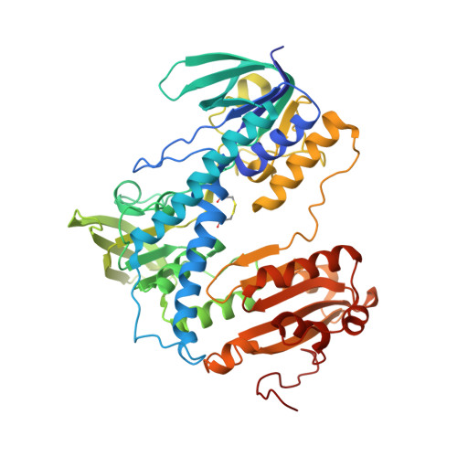
    Selenoproteins contain a highly reactive 21st amino acid selenocysteine (Sec) encoded by recoding of a predefined UGA codon. Because of a lack of selenoprotein supply, high chemical reactivity of Sec, and intricate translation machineries, selenoprotein crystal structures are difficult to obtain. Structural prerequisites for Sec involvement in enzyme catalysis are therefore sparsely known. Here we present the crystal structure of catalytically active rat thioredoxin reductase 1 (TrxR1), revealing surprises at the C-terminal Sec-containing active site in view of previous literature. The oxidized enzyme presents a selenenylsulfide motif in trans-configuration, with the selenium atom of Sec-498 positioned beneath the side chain of Tyr-116, thereby located far from the redox active moieties proposed to be involved in electron transport to the Sec-containing active site. Upon reduction to a selenolthiol motif, the Sec residue moved toward solvent exposure, consistent with its presumed role in reduction of TrxR1 substrates or as target of electrophilic agents inhibiting the enzyme. A Y116I mutation lowered catalytic efficiency in reduction of thioredoxin, but surprisingly increased turnover using 5-hydroxy-1,4-naphthoquinone (juglone) as substrate. The same mutation also decreased sensitivity to inhibition by cisplatin. The results suggest that Tyr-116 plays an important role for catalysis of TrxR1 by interacting with the selenenylsulfide of oxidized TrxR1, thereby facilitating its reduction in the reductive half-reaction of the enzyme. The interaction of a selenenylsulfide with the phenyl ring of a tyrosine, affecting turnover, switch of substrate specificity, and modulation of sensitivity to electrophilic agents, gives important clues into the mechanism of TrxR1, which is a selenoprotein that plays a major role for mammalian cell fate and function. The results also demonstrate that a recombinant selenoprotein TrxR can be produced in high amount and sufficient purity to enable crystal structure determination, which suggests that additional structural studies of these types of proteins are feasible.


  • Organizational Affiliation

    Division of Biochemistry, Medical Nobel Institute for Biochemistry, Karolinska Institutet, SE-171 77 Stockholm, Sweden.


Macromolecules
Find similar proteins by:  (by identity cutoff)  |  3D Structure
Entity ID: 1
MoleculeChains Sequence LengthOrganismDetailsImage
Thioredoxin reductase 1, cytoplasmic
A, B, C, D, E
A, B, C, D, E, F
499Rattus norvegicusMutation(s): 0 
Gene Names: Txnrd1Trxr1
EC: 1.8.1.9
UniProt
Find proteins for O89049 (Rattus norvegicus)
Explore O89049 
Go to UniProtKB:  O89049
Entity Groups  
Sequence Clusters30% Identity50% Identity70% Identity90% Identity95% Identity100% Identity
UniProt GroupO89049
Sequence Annotations
Expand
  • Reference Sequence
Small Molecules
Ligands 2 Unique
IDChains Name / Formula / InChI Key2D Diagram3D Interactions
FAD
Query on FAD

Download Ideal Coordinates CCD File 
G [auth A]
I [auth B]
K [auth C]
M [auth D]
O [auth E]
G [auth A],
I [auth B],
K [auth C],
M [auth D],
O [auth E],
Q [auth F]
FLAVIN-ADENINE DINUCLEOTIDE
C27 H33 N9 O15 P2
VWWQXMAJTJZDQX-UYBVJOGSSA-N
NAP
Query on NAP

Download Ideal Coordinates CCD File 
H [auth A]
J [auth B]
L [auth C]
N [auth D]
P [auth E]
H [auth A],
J [auth B],
L [auth C],
N [auth D],
P [auth E],
R [auth F]
NADP NICOTINAMIDE-ADENINE-DINUCLEOTIDE PHOSPHATE
C21 H28 N7 O17 P3
XJLXINKUBYWONI-NNYOXOHSSA-N
Experimental Data & Validation

Experimental Data

  • Method: X-RAY DIFFRACTION
  • Resolution: 3.10 Å
  • R-Value Free: 0.289 
  • R-Value Work: 0.256 
  • R-Value Observed: 0.258 
  • Space Group: P 1 21 1
Unit Cell:
Length ( Å )Angle ( ˚ )
a = 78.231α = 90
b = 137.754β = 94
c = 168.954γ = 90
Software Package:
Software NamePurpose
ProDCdata collection
MOLREPphasing
REFMACrefinement
MOSFLMdata reduction
SCALAdata scaling

Structure Validation

View Full Validation Report


Ligand Structure Quality Assessment 


Entry History 

Deposition Data

Revision History  (Full details and data files)

  • Version 1.0: 2008-12-02
    Type: Initial release
  • Version 1.1: 2011-07-13
    Changes: Version format compliance
  • Version 1.2: 2012-08-29
    Changes: Derived calculations
  • Version 1.3: 2014-02-19
    Changes: Atomic model, Database references, Structure summary
  • Version 1.4: 2014-08-06
    Changes: Derived calculations, Structure summary
  • Version 1.5: 2018-03-07
    Changes: Data collection
  • Version 1.6: 2023-08-30
    Changes: Data collection, Database references, Derived calculations, Refinement description