3DA0

Crystal structure of a cleaved form of a chimeric receptor binding protein from Lactococcal phages subspecies TP901-1 and p2


Experimental Data Snapshot

  • Method: X-RAY DIFFRACTION
  • Resolution: 1.65 Å
  • R-Value Free: 0.201 
  • R-Value Work: 0.164 
  • R-Value Observed: 0.165 

wwPDB Validation   3D Report Full Report


This is version 1.3 of the entry. See complete history


Literature

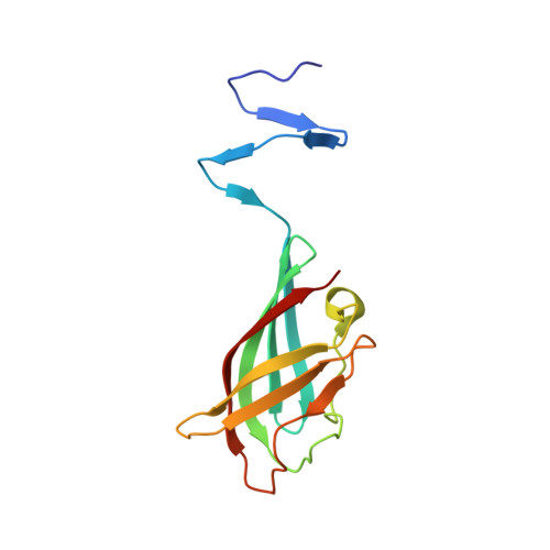
Crystal structure of a chimeric receptor binding protein constructed from two lactococcal phages.

Siponen, M.Spinelli, S.Blangy, S.Moineau, S.Cambillau, C.Campanacci, V.

(2009) J Bacteriol 191: 3220-3225

  • DOI: https://doi.org/10.1128/JB.01637-08
  • Primary Citation of Related Structures:  
    3D8M, 3DA0

  • PubMed Abstract: 

    Lactococcus lactis, a gram-positive bacterium widely used by the dairy industry to manufacture cheeses, is subject to infection by a diverse population of virulent phages. We have previously determined the structures of three receptor binding proteins (RBPs) from lactococcal phages TP901-1, p2, and bIL170, each of them having a distinct host range. Virulent phages p2 and bIL170 are classified within the 936 group, while the temperate phage TP901-1 is a member of the genetically distinct P335 polythetic group. These RBPs comprise three domains: the N-terminal domain, binding to the virion particle; a beta-helical linker domain; and the C-terminal domain, bearing the receptor binding site used for host recognition. Here, we have designed, expressed, and determined the structure of an RBP chimera in which the N-terminal and linker RBP domains of phage TP901-1 (P335) are fused to the C-terminal RBP domain of phage p2 (936). This chimera exhibits a stable structure that closely resembles the parental structures, while a slight displacement of the linker made RBP domain adaptation efficient. The receptor binding site is structurally indistinguishable from that of native p2 RBP and binds glycerol with excellent affinity.


  • Organizational Affiliation

    Architecture et Fonction des Macromolécules Biologiques, UMR 6098 CNRS, and Universités Aix-Marseille I & II, Campus de Luminy, Case 932, 13288 Marseille Cedex 09, France.


Macromolecules
Find similar proteins by:  (by identity cutoff)  |  3D Structure
Entity ID: 1
MoleculeChains Sequence LengthOrganismDetailsImage
Cleaved chimeric receptor binding protein from bacteriophages TP901-1 and p2
A, B, C
136Lactococcus phage TP901-1Lactococcus virus P2
This entity is chimeric
Mutation(s): 0 
Gene Names: bpprbp
UniProt
Find proteins for Q9G096 (Lactococcus phage TP901-1)
Explore Q9G096 
Go to UniProtKB:  Q9G096
Find proteins for Q71AW2 (Lactococcus phage p2)
Explore Q71AW2 
Go to UniProtKB:  Q71AW2
Entity Groups  
Sequence Clusters30% Identity50% Identity70% Identity90% Identity95% Identity100% Identity
UniProt GroupsQ9G096Q71AW2
Sequence Annotations
Expand
  • Reference Sequence
Experimental Data & Validation

Experimental Data

  • Method: X-RAY DIFFRACTION
  • Resolution: 1.65 Å
  • R-Value Free: 0.201 
  • R-Value Work: 0.164 
  • R-Value Observed: 0.165 
  • Space Group: P 1 21 1
Unit Cell:
Length ( Å )Angle ( ˚ )
a = 57.168α = 90
b = 43.682β = 101.63
c = 78.946γ = 90
Software Package:
Software NamePurpose
REFMACrefinement
ADSCdata collection
MOSFLMdata reduction
SCALAdata scaling
PHASERphasing

Structure Validation

View Full Validation Report


Entry History 

Deposition Data

Revision History  (Full details and data files)

  • Version 1.0: 2009-06-09
    Type: Initial release
  • Version 1.1: 2011-07-13
    Changes: Advisory, Version format compliance
  • Version 1.2: 2017-06-21
    Changes: Database references, Source and taxonomy
  • Version 1.3: 2023-08-30
    Changes: Data collection, Database references, Refinement description