3CK6

Crystal structure of ZntB cytoplasmic domain from Vibrio parahaemolyticus RIMD 2210633


Experimental Data Snapshot

  • Method: X-RAY DIFFRACTION
  • Resolution: 1.90 Å
  • R-Value Free: 0.223 
  • R-Value Work: 0.185 
  • R-Value Observed: 0.187 

wwPDB Validation   3D Report Full Report


This is version 1.2 of the entry. See complete history


Literature

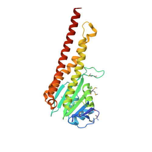
Structure and electrostatic property of cytoplasmic domain of ZntB transporter.

Tan, K.Sather, A.Robertson, J.L.Moy, S.Roux, B.Joachimiak, A.

(2009) Protein Sci 18: 2043-2052

  • DOI: https://doi.org/10.1002/pro.215
  • Primary Citation of Related Structures:  
    3CK6

  • PubMed Abstract: 

    ZntB is the distant homolog of CorA Mg(2+) transporter within the metal ion transporter superfamily. It was early reported that the ZntB from Salmonella typhimurium facilitated efflux of Zn(2+) and Cd(2+), but not Mg(2+). Here, we report the 1.90 A crystal structure of the intracellular domain of ZntB from Vibrio parahemolyticus. The domain forms a funnel-shaped homopentamer that is similar to the full-length CorA from Thermatoga maritima, but differs from two previously reported dimeric structures of truncated CorA intracellular domains. However, no Zn(2+) or Cd(2+) binding sites were identified in the high-resolution structure. Instead, 25 well-defined Cl(-) ions were observed and some of these binding sites are highly conserved within the ZntB family. Continuum electrostatics calculations suggest that the central pore of the funnel is highly attractive for cations, especially divalents. The presence of the bound Cl(-) ions increases the stability of cations along the pore suggesting they could be important in enhancing cation transport.


  • Organizational Affiliation

    Midwest Center for Structural Genomics and Structural Biology Center, Argonne, Illinois 60439, USA.


Macromolecules
Find similar proteins by:  (by identity cutoff)  |  3D Structure
Entity ID: 1
MoleculeChains Sequence LengthOrganismDetailsImage
Putative membrane transport protein
A, B, C, D, E
252Vibrio parahaemolyticus RIMD 2210633Mutation(s): 1 
Gene Names: VP2389NP_798768.1
Membrane Entity: Yes 
UniProt
Find proteins for Q87M69 (Vibrio parahaemolyticus serotype O3:K6 (strain RIMD 2210633))
Explore Q87M69 
Go to UniProtKB:  Q87M69
Entity Groups  
Sequence Clusters30% Identity50% Identity70% Identity90% Identity95% Identity100% Identity
UniProt GroupQ87M69
Sequence Annotations
Expand
  • Reference Sequence
Small Molecules
Ligands 1 Unique
IDChains Name / Formula / InChI Key2D Diagram3D Interactions
CL
Query on CL

Download Ideal Coordinates CCD File 
AA [auth E]
BA [auth E]
CA [auth E]
DA [auth E]
F [auth A]
AA [auth E],
BA [auth E],
CA [auth E],
DA [auth E],
F [auth A],
G [auth A],
H [auth A],
I [auth A],
J [auth A],
K [auth B],
L [auth B],
M [auth B],
N [auth B],
O [auth B],
P [auth C],
Q [auth C],
R [auth C],
S [auth C],
T [auth C],
U [auth D],
V [auth D],
W [auth D],
X [auth D],
Y [auth D],
Z [auth E]
CHLORIDE ION
Cl
VEXZGXHMUGYJMC-UHFFFAOYSA-M
Modified Residues  1 Unique
IDChains TypeFormula2D DiagramParent
MSE
Query on MSE
A, B, C, D, E
L-PEPTIDE LINKINGC5 H11 N O2 SeMET
Experimental Data & Validation

Experimental Data

  • Method: X-RAY DIFFRACTION
  • Resolution: 1.90 Å
  • R-Value Free: 0.223 
  • R-Value Work: 0.185 
  • R-Value Observed: 0.187 
  • Space Group: P 1 21 1
Unit Cell:
Length ( Å )Angle ( ˚ )
a = 64.39α = 90
b = 130.213β = 103.41
c = 79.386γ = 90
Software Package:
Software NamePurpose
REFMACrefinement
SBC-Collectdata collection
HKL-3000data reduction
HKL-3000data scaling
SHELXDphasing
MLPHAREphasing
DMphasing
RESOLVEphasing
HKL-3000phasing

Structure Validation

View Full Validation Report


Entry History 

Deposition Data

Revision History  (Full details and data files)

  • Version 1.0: 2008-03-25
    Type: Initial release
  • Version 1.1: 2011-07-13
    Changes: Advisory, Version format compliance
  • Version 1.2: 2021-10-20
    Changes: Database references, Derived calculations