The Catalytic Activity of Protein-disulfide Isomerase Requires a Conformationally Flexible Molecule.
Tian, G., Kober, F.X., Lewandrowski, U., Sickmann, A., Lennarz, W.J., Schindelin, H.(2008) J Biol Chem 283: 33630-33640
- PubMed: 18815132
- DOI: https://doi.org/10.1074/jbc.M806026200
- Primary Citation of Related Structures:
3BOA - PubMed Abstract:
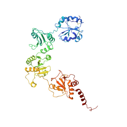
Protein-disulfide isomerase (PDI) catalyzes the formation of the correct pattern of disulfide bonds in secretory proteins. A low resolution crystal structure of yeast PDI described here reveals large scale conformational changes compared with the initially reported structure, indicating that PDI is a highly flexible molecule with its catalytic domains, a and a', representing two mobile arms connected to a more rigid core composed of the b and b' domains. Limited proteolysis revealed that the linker between the a domain and the core is more susceptible to degradation than that connecting the a' domain to the core. By restricting the two arms with inter-domain disulfide bonds, the molecular flexibility of PDI, especially that of its a domain, was demonstrated to be essential for the enzymatic activity in vitro and in vivo. The crystal structure also featured a PDI dimer, and a propensity to dimerize in solution and in the ER was confirmed by cross-linking experiments and the split green fluorescent protein system. Although sedimentation studies suggested that the self-association of PDI is weak, we hypothesize that PDI exists as an interconvertible mixture of monomers and dimers in the endoplasmic reticulum due to its high abundance in this compartment.
Organizational Affiliation:
Department of Biochemistry and Cell Biology, Stony Brook University, Stony Brook, New York 11794-5215, USA.