3BJX

Structure of a Group I haloacid dehalogenase from Pseudomonas putida strain PP3


Experimental Data Snapshot

  • Method: X-RAY DIFFRACTION
  • Resolution: 2.30 Å
  • R-Value Free: 0.250 
  • R-Value Work: 0.181 
  • R-Value Observed: 0.185 

wwPDB Validation   3D Report Full Report


This is version 1.1 of the entry. See complete history


Literature

The crystal structure of DehI reveals a new alpha-haloacid dehalogenase fold and active-site mechanism

Schmidberger, J.W.Wilce, J.A.Weightman, A.J.Whisstock, J.C.Wilce, M.C.J.

(2008) J Mol Biol 378: 284-294

  • DOI: https://doi.org/10.1016/j.jmb.2008.02.035
  • Primary Citation of Related Structures:  
    3BJX

  • PubMed Abstract: 

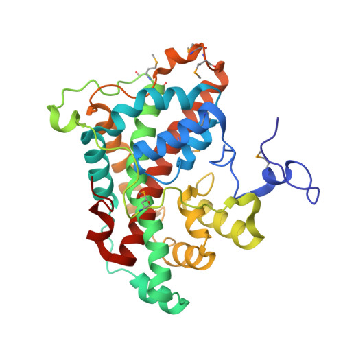
    Haloacid dehalogenases catalyse the removal of halides from organic haloacids and are of interest for bioremediation and for their potential use in the synthesis of industrial chemicals. We present the crystal structure of the homodimer DehI from Pseudomonas putida strain PP3, the first structure of a group I alpha-haloacid dehalogenase that can process both L- and D-substrates. The structure shows that the DehI monomer consists of two domains of approximately 130 amino acids that have approximately 16% sequence identity yet adopt virtually identical and unique folds that form a pseudo-dimer. Analysis of the active site reveals the likely binding mode of both L- and D-substrates with respect to key catalytic residues. Asp189 is predicted to activate a water molecule for nucleophilic attack of the substrate chiral centre resulting in an inversion of configuration of either l- or d-substrates in contrast to D-only enzymes. These details will assist with future bioengineering of dehalogenases.


  • Organizational Affiliation

    Department of Pharmacology, School of Medicine and Pharmacology, The University of Western Australia, Perth, Western Australia 6907, Australia.


Macromolecules
Find similar proteins by:  (by identity cutoff)  |  3D Structure
Entity ID: 1
MoleculeChains Sequence LengthOrganismDetailsImage
Halocarboxylic acid dehalogenase DehI
A, B, C, D
311Pseudomonas putidaMutation(s): 0 
Gene Names: dehI
EC: 3.8.1.2
UniProt
Find proteins for Q8GJ84 (Pseudomonas putida)
Explore Q8GJ84 
Go to UniProtKB:  Q8GJ84
Entity Groups  
Sequence Clusters30% Identity50% Identity70% Identity90% Identity95% Identity100% Identity
UniProt GroupQ8GJ84
Sequence Annotations
Expand
  • Reference Sequence
Small Molecules
Ligands 1 Unique
IDChains Name / Formula / InChI Key2D Diagram3D Interactions
SO4
Query on SO4

Download Ideal Coordinates CCD File 
E [auth A]
F [auth A]
G [auth A]
H [auth A]
I [auth B]
E [auth A],
F [auth A],
G [auth A],
H [auth A],
I [auth B],
J [auth B],
K [auth B],
L [auth C],
M [auth C],
N [auth C],
O [auth C],
P [auth D],
Q [auth D],
R [auth D],
S [auth D],
T [auth D]
SULFATE ION
O4 S
QAOWNCQODCNURD-UHFFFAOYSA-L
Modified Residues  1 Unique
IDChains TypeFormula2D DiagramParent
MSE
Query on MSE
A, B, C, D
L-PEPTIDE LINKINGC5 H11 N O2 SeMET
Experimental Data & Validation

Experimental Data

  • Method: X-RAY DIFFRACTION
  • Resolution: 2.30 Å
  • R-Value Free: 0.250 
  • R-Value Work: 0.181 
  • R-Value Observed: 0.185 
  • Space Group: P 1 21 1
Unit Cell:
Length ( Å )Angle ( ˚ )
a = 68.323α = 90
b = 111.864β = 93.71
c = 75.181γ = 90
Software Package:
Software NamePurpose
MOSFLMdata reduction
SCALAdata scaling
SOLVEphasing
RESOLVEphasing
REFMACrefinement
PDB_EXTRACTdata extraction
CrystalCleardata collection

Structure Validation

View Full Validation Report


Entry History 

Deposition Data

Revision History  (Full details and data files)

  • Version 1.0: 2008-04-01
    Type: Initial release
  • Version 1.1: 2011-07-13
    Changes: Version format compliance