Alternative conformations of the x region of human protein disulphide-isomerase modulate exposure of the substrate binding b' domain
Nguyen, V.D., Wallis, K., Howard, M.J., Haapalainen, A.M., Salo, K.E.H., Saaranen, M.J., Sidhu, A., Wierenga, R.K., Freedman, R.B., Ruddock, L.W., Williamson, R.A.(2008) J Mol Biol 383: 1144-1155
- PubMed: 18801374
- DOI: https://doi.org/10.1016/j.jmb.2008.08.085
- Primary Citation of Related Structures:
3BJ5 - PubMed Abstract:
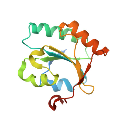
Protein disulphide isomerase (PDI) is a key multi-domain protein folding catalyst in the endoplasmic reticulum. The b' domain of PDI is essential for the non-covalent binding of incompletely folded protein substrates. Earlier, we defined the substrate binding site in the b' domain of human PDI by modelling and mutagenesis studies. Here, we show by fluorescence and NMR that recombinant human PDI b'x (comprising the b' domain and the subsequent x linker region) can assume at least two different conformations in solution. We have screened mutants in the b'x region to identify mutations that favour one of these conformers in recombinant b'x, and isolated and characterised examples of both types. We have crystallised one mutant of b'x (I272A mutation) in which one conformer is stabilized, and determined its crystal structure to a resolution of 2.2 A. This structure shows that the b' domain has the typical thioredoxin fold and that the x region can interact with the b' domain by "capping" a hydrophobic site on the b' domain. This site is most likely the substrate binding site and hence such capping will inhibit substrate binding. All of the mutations we previously reported to inhibit substrate binding shift the equilibrium towards the capped conformer. Hence, these mutations act by altering the natural equilibrium and decreasing the accessibility of the substrate binding site. Furthermore, we have confirmed that the corresponding structural transition occurs in the wild type full-length PDI. A cross-comparison of our data with that for other PDI-family members, Pdi1p and ERp44, suggests that the x region of PDI can adopt alternative conformations during the functional cycle of PDI action and that these are linked to the ability of PDI to interact with folding substrates.
Organizational Affiliation:
Department of Biochemistry, University of Oulu, FIN-90014 Oulu, Finland.