


Crystal Structure of an Acetylacetone Dioxygenase from Acinetobacter johnsonii
Stranzl, G.R., Wagner, U.G., Straganz, G., Steiner, W., Kratky, C.To be published.
Experimental Data Snapshot
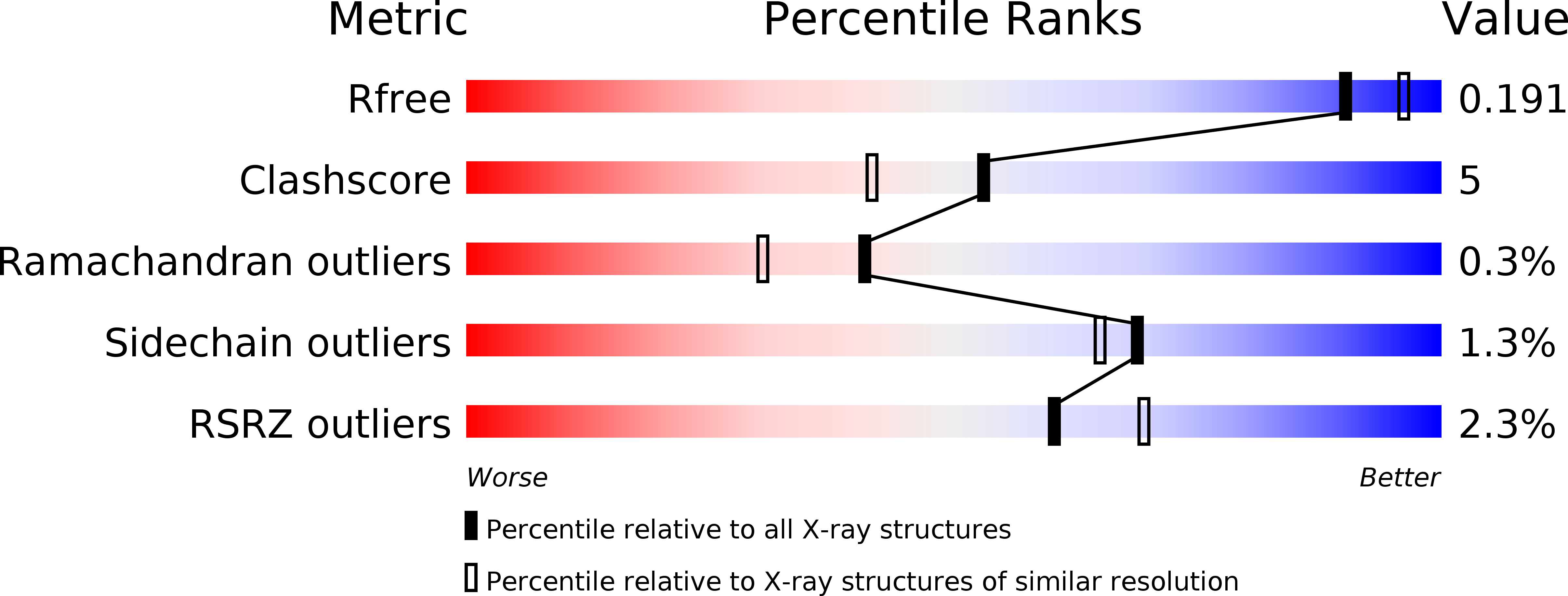
wwPDB Validation 3D Report Full Report
Entity ID: 1 | |||||
|---|---|---|---|---|---|
| Molecule | Chains | Sequence Length | Organism | Details | Image |
| Acetylacetone-cleaving enzyme | 153 | Acinetobacter johnsonii | Mutation(s): 0 EC: 1.13.11.50 | 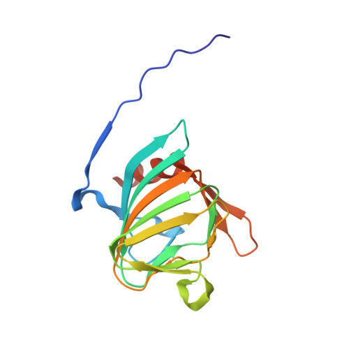 | |
UniProt | |||||
Find proteins for Q8GNT2 (Acinetobacter johnsonii) Explore Q8GNT2 Go to UniProtKB: Q8GNT2 | |||||
Entity Groups | |||||
| Sequence Clusters | 30% Identity50% Identity70% Identity90% Identity95% Identity100% Identity | ||||
| UniProt Group | Q8GNT2 | ||||
Sequence AnnotationsExpand | |||||
| |||||
| Ligands 1 Unique | |||||
|---|---|---|---|---|---|
| ID | Chains | Name / Formula / InChI Key | 2D Diagram | 3D Interactions | |
| ZN Query on ZN | E [auth A], F [auth B], G [auth C], H [auth D] | ZINC ION Zn PTFCDOFLOPIGGS-UHFFFAOYSA-N |  | ||
| Length ( Å ) | Angle ( ˚ ) |
|---|---|
| a = 43.2 | α = 69.85 |
| b = 57.2 | β = 74.32 |
| c = 62.93 | γ = 69.71 |
| Software Name | Purpose |
|---|---|
| DENZO | data reduction |
| SCALEPACK | data scaling |
| SOLVE | phasing |
| RESOLVE | phasing |
| CNS | refinement |
| PDB_EXTRACT | data extraction |
| MAR345 | data collection |
| HKL-2000 | data reduction |
| HKL-2000 | data scaling |
RCSB PDB (citation) is hosted by
RCSB PDB is a member of the

