Structure of a novel phosphotyrosine-binding domain in Hakai that targets E-cadherin
Mukherjee, M., Chow, S.Y., Yusoff, P., Seetharaman, J., Ng, C., Sinniah, S., Koh, X.W., Asgar, N.F., Li, D., Yim, D., Jackson, R.A., Yew, J., Qian, J., Iyu, A., Lim, Y.P., Zhou, X., Sze, S.K., Guy, G.R., Sivaraman, J.(2012) EMBO J 31: 1308-1319
- PubMed: 22252131
- DOI: https://doi.org/10.1038/emboj.2011.496
- Primary Citation of Related Structures:
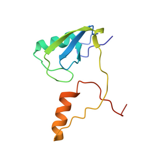
3VK6 - PubMed Abstract:
Phosphotyrosine-binding domains, typified by the SH2 (Src homology 2) and PTB domains, are critical upstream components of signal transduction pathways. The E3 ubiquitin ligase Hakai targets tyrosine-phosphorylated E-cadherin via an uncharacterized domain. In this study, the crystal structure of Hakai (amino acids 106-206) revealed that it forms an atypical, zinc-coordinated homodimer by utilizing residues from the phosphotyrosine-binding domain of two Hakai monomers. Hakai dimerization allows the formation of a phosphotyrosine-binding pocket that recognizes specific phosphorylated tyrosines and flanking acidic amino acids of Src substrates, such as E-cadherin, cortactin and DOK1. NMR and mutational analysis identified the Hakai residues required for target binding within the binding pocket, now named the HYB domain. ZNF645 also possesses a HYB domain but demonstrates different target specificities. The HYB domain is structurally different from other phosphotyrosine-binding domains and is a potential drug target due to its novel structural features.
- Department of Biological Sciences, National University of Singapore, Singapore.
Organizational Affiliation: