3UGU

Crystal Structure of p44 (Splice Variant of Visual Arrestin)


Experimental Data Snapshot

  • Method: X-RAY DIFFRACTION
  • Resolution: 1.85 Å
  • R-Value Free: 0.214 
  • R-Value Work: 0.174 
  • R-Value Observed: 0.176 

wwPDB Validation   3D Report Full Report


This is version 1.3 of the entry. See complete history


Literature

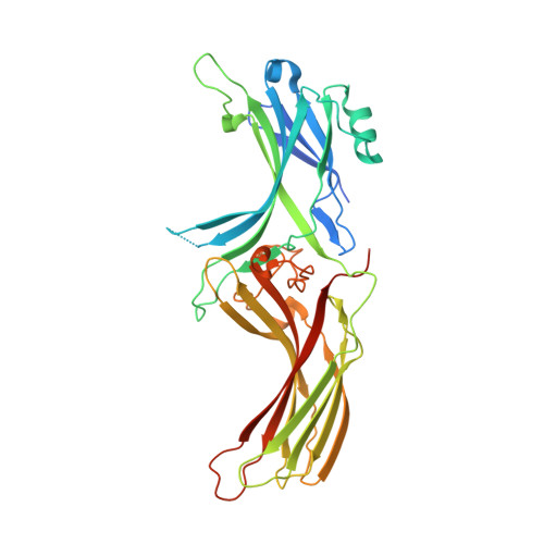
Crystal Structure of p44, a Constitutively Active Splice Variant of Visual Arrestin.

Granzin, J.Cousin, A.Weirauch, M.Schlesinger, R.Buldt, G.Batra-Safferling, R.

(2012) J Mol Biol 416: 611-618

  • DOI: https://doi.org/10.1016/j.jmb.2012.01.028
  • Primary Citation of Related Structures:  
    3UGU, 3UGX

  • PubMed Abstract: 

    Visual arrestin specifically binds to photoactivated and phosphorylated rhodopsin and inactivates phototransduction. In contrast, the p44 splice variant can terminate phototransduction by binding to nonphosphorylated light-activated rhodopsin. Here we report the crystal structure of bovine p44 at a resolution of 1.85 Å. Compared to native arrestin, the p44 structure reveals significant differences in regions crucial for receptor binding, namely flexible loop V-VI and polar core regions. Additionally, electrostatic potential is remarkably positive on the N-domain and the C-domain. The p44 structure represents an active conformation that serves as a model to explain the 'constitutive activity' found in arrestin variants.


  • Organizational Affiliation

    Institute of Complex Systems, ICS-6: Structural Biochemistry, Forschungszentrum Jülich, 52425 Jülich, Germany.


Macromolecules
Find similar proteins by:  (by identity cutoff)  |  3D Structure
Entity ID: 1
MoleculeChains Sequence LengthOrganismDetailsImage
S-arrestin380Bos taurusMutation(s): 0 
Gene Names: SAG
UniProt
Find proteins for P08168 (Bos taurus)
Explore P08168 
Go to UniProtKB:  P08168
Entity Groups  
Sequence Clusters30% Identity50% Identity70% Identity90% Identity95% Identity100% Identity
UniProt GroupP08168
Sequence Annotations
Expand
  • Reference Sequence
Experimental Data & Validation

Experimental Data

  • Method: X-RAY DIFFRACTION
  • Resolution: 1.85 Å
  • R-Value Free: 0.214 
  • R-Value Work: 0.174 
  • R-Value Observed: 0.176 
  • Space Group: P 21 21 21
Unit Cell:
Length ( Å )Angle ( ˚ )
a = 66.964α = 90
b = 71.583β = 90
c = 79.7γ = 90
Software Package:
Software NamePurpose
ADSCdata collection
MOLREPphasing
PHENIXrefinement
MOSFLMdata reduction
SCALAdata scaling

Structure Validation

View Full Validation Report


Entry History 

Deposition Data

Revision History  (Full details and data files)

  • Version 1.0: 2012-02-08
    Type: Initial release
  • Version 1.1: 2012-02-15
    Changes: Structure summary
  • Version 1.2: 2012-03-21
    Changes: Database references
  • Version 1.3: 2024-02-28
    Changes: Data collection, Database references