3RVD

Crystal structure of the binary complex, obtained by soaking, of photosyntetic a4 glyceraldehyde 3-phosphate dehydrogenase (gapdh) with cp12-2, both from arabidopsis thaliana.


Experimental Data Snapshot

  • Method: X-RAY DIFFRACTION
  • Resolution: 2.70 Å
  • R-Value Free: 0.318 
  • R-Value Work: 0.247 
  • R-Value Observed: 0.247 

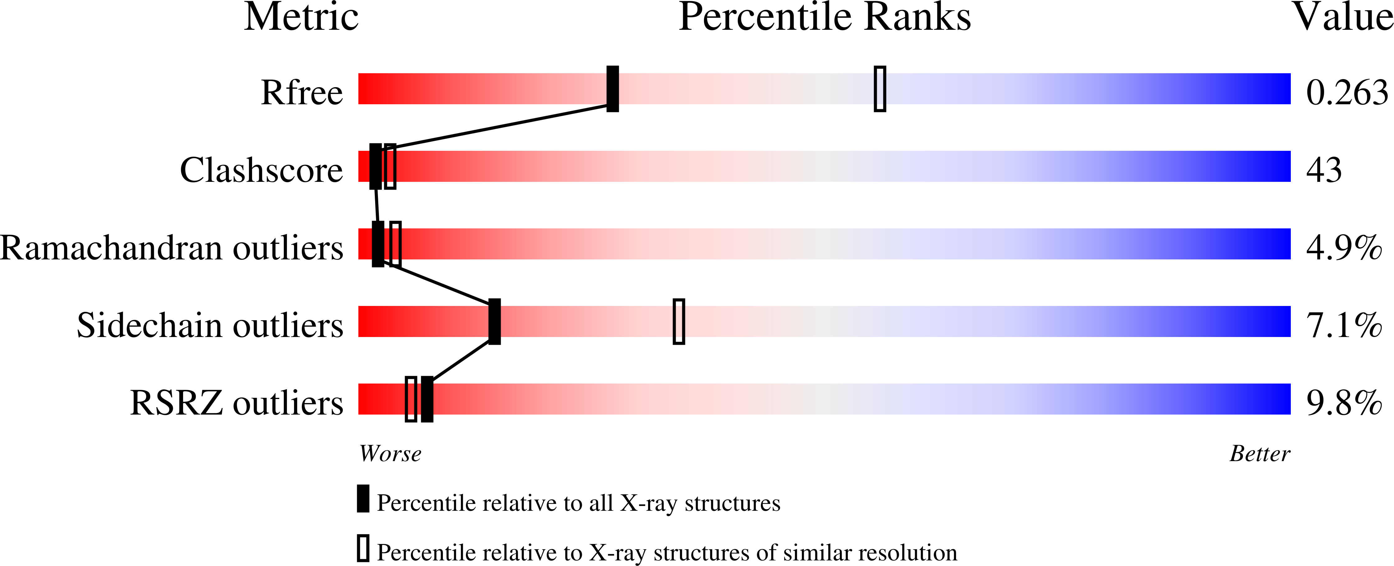
wwPDB Validation   3D Report Full Report


Ligand Structure Quality Assessment 


This is version 1.4 of the entry. See complete history


Literature

Conformational Selection and Folding-upon-binding of Intrinsically Disordered Protein CP12 Regulate Photosynthetic Enzymes Assembly.

Fermani, S.Trivelli, X.Sparla, F.Thumiger, A.Calvaresi, M.Marri, L.Falini, G.Zerbetto, F.Trost, P.

(2012) J Biol Chem 287: 21372-21383

  • DOI: https://doi.org/10.1074/jbc.M112.350355
  • Primary Citation of Related Structures:  
    2LJ9, 3QV1, 3RVD

  • PubMed Abstract: 

    Carbon assimilation in plants is regulated by the reduction of specific protein disulfides by light and their re-oxidation in the dark. The redox switch CP12 is an intrinsically disordered protein that can form two disulfide bridges. In the dark oxidized CP12 forms an inactive supramolecular complex with glyceraldehyde-3-phosphate dehydrogenase (GAPDH) and phosphoribulokinase, two enzymes of the carbon assimilation cycle. Here we show that binding of CP12 to GAPDH, the first step of ternary complex formation, follows an integrated mechanism that combines conformational selection with induced folding steps. Initially, a CP12 conformation characterized by a circular structural motif including the C-terminal disulfide is selected by GAPDH. Subsequently, the induced folding of the flexible C-terminal tail of CP12 in the active site of GAPDH stabilizes the binary complex. Formation of several hydrogen bonds compensates the entropic cost of CP12 fixation and terminates the interaction mechanism that contributes to carbon assimilation control.


  • Organizational Affiliation

    Department of Chemistry G Ciamician, University of Bologna, 40126 Bologna, Italy.


Macromolecules
Find similar proteins by:  (by identity cutoff)  |  3D Structure
Entity ID: 1
MoleculeChains Sequence LengthOrganismDetailsImage
Glyceraldehyde-3-phosphate dehydrogenase A, chloroplastic
A,
B,
C,
D,
E,
F,
G,
H,
O,
P [auth Q]
337Arabidopsis thalianaMutation(s): 0 
Gene Names: At3g26650GAPAMLJ15.4MLJ15_5
EC: 1.2.1.13
UniProt
Find proteins for P25856 (Arabidopsis thaliana)
Explore P25856 
Go to UniProtKB:  P25856
Entity Groups  
Sequence Clusters30% Identity50% Identity70% Identity90% Identity95% Identity100% Identity
UniProt GroupP25856
Sequence Annotations
Expand
  • Reference Sequence
Find similar proteins by:  (by identity cutoff)  |  3D Structure
Entity ID: 2
MoleculeChains Sequence LengthOrganismDetailsImage
Photosyntetic glyceraldehyde-3-phosphate dehydrogenase (a4 isoform)
I, J, K, L, M
I, J, K, L, M, N
82Arabidopsis thalianaMutation(s): 0 
Gene Names: At3g26650
UniProt
Find proteins for Q9LZP9 (Arabidopsis thaliana)
Explore Q9LZP9 
Go to UniProtKB:  Q9LZP9
Entity Groups  
Sequence Clusters30% Identity50% Identity70% Identity90% Identity95% Identity100% Identity
UniProt GroupQ9LZP9
Sequence Annotations
Expand
  • Reference Sequence
Small Molecules
Ligands 2 Unique
IDChains Name / Formula / InChI Key2D Diagram3D Interactions
NAD
Query on NAD

Download Ideal Coordinates CCD File 
BA [auth C]
HA [auth D]
JA [auth E]
NA [auth F]
Q [auth A]
BA [auth C],
HA [auth D],
JA [auth E],
NA [auth F],
Q [auth A],
QA [auth G],
SA [auth H],
UA [auth O],
XA [auth Q],
Y [auth B]
NICOTINAMIDE-ADENINE-DINUCLEOTIDE
C21 H27 N7 O14 P2
BAWFJGJZGIEFAR-NNYOXOHSSA-N
SO4
Query on SO4

Download Ideal Coordinates CCD File 
AA [auth B]
AB [auth Q]
BB [auth Q]
CA [auth C]
DA [auth C]
AA [auth B],
AB [auth Q],
BB [auth Q],
CA [auth C],
DA [auth C],
EA [auth C],
FA [auth C],
GA [auth C],
IA [auth D],
KA [auth E],
LA [auth E],
MA [auth E],
OA [auth F],
PA [auth F],
R [auth A],
RA [auth G],
S [auth A],
T [auth A],
TA [auth H],
U [auth A],
V [auth A],
VA [auth O],
W [auth A],
WA [auth O],
X [auth A],
YA [auth Q],
Z [auth B],
ZA [auth Q]
SULFATE ION
O4 S
QAOWNCQODCNURD-UHFFFAOYSA-L
Experimental Data & Validation

Experimental Data

  • Method: X-RAY DIFFRACTION
  • Resolution: 2.70 Å
  • R-Value Free: 0.318 
  • R-Value Work: 0.247 
  • R-Value Observed: 0.247 
  • Space Group: I 2 2 2
Unit Cell:
Length ( Å )Angle ( ˚ )
a = 153.183α = 90
b = 188.75β = 90
c = 312.17γ = 90
Software Package:
Software NamePurpose
ADSCdata collection
AMoREphasing
CNSrefinement
DENZOdata reduction
SCALEPACKdata scaling

Structure Validation

View Full Validation Report


Ligand Structure Quality Assessment 


Entry History 

Deposition Data

Revision History  (Full details and data files)

  • Version 1.0: 2012-04-25
    Type: Initial release
  • Version 1.1: 2012-07-04
    Changes: Database references
  • Version 1.2: 2014-11-12
    Changes: Structure summary
  • Version 1.3: 2023-09-13
    Changes: Data collection, Database references, Derived calculations, Refinement description
  • Version 1.4: 2024-03-13
    Changes: Source and taxonomy