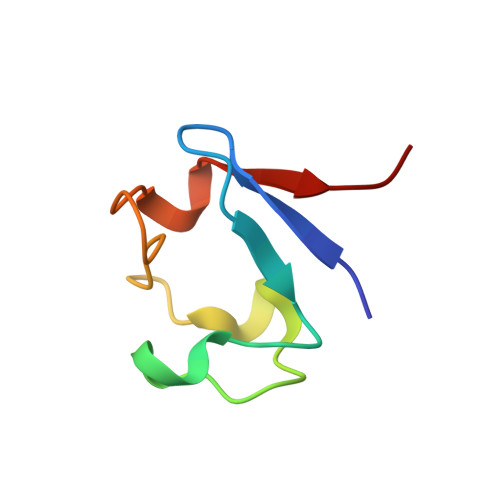
Unambiguous determination of H-atom positions: comparing results from neutron and high-resolution X-ray crystallography.
Gardberg, A.S., Del Castillo, A.R., Weiss, K.L., Meilleur, F., Blakeley, M.P., Myles, D.A.(2010) Acta Crystallogr D Biol Crystallogr 66: 558-567
- PubMed: 20445231
- DOI: https://doi.org/10.1107/S0907444910005494
- Primary Citation of Related Structures:
3KYU, 3KYV, 3KYW, 3KYX, 3KYY - PubMed Abstract:
The locations of H atoms in biological structures can be difficult to determine using X-ray diffraction methods. Neutron diffraction offers a relatively greater scattering magnitude from H and D atoms. Here, 1.65 A resolution neutron diffraction studies of fully perdeuterated and selectively CH(3)-protonated perdeuterated crystals of Pyrococcus furiosus rubredoxin (D-rubredoxin and HD-rubredoxin, respectively) at room temperature (RT) are described, as well as 1.1 A resolution X-ray diffraction studies of the same protein at both RT and 100 K. The two techniques are quantitatively compared in terms of their power to directly provide atomic positions for D atoms and analyze the role played by atomic thermal motion by computing the sigma level at the D-atom coordinate in simulated-annealing composite D-OMIT maps. It is shown that 1.65 A resolution RT neutron data for perdeuterated rubredoxin are approximately 8 times more likely overall to provide high-confidence positions for D atoms than 1.1 A resolution X-ray data at 100 K or RT. At or above the 1.0sigma level, the joint X-ray/neutron (XN) structures define 342/378 (90%) and 291/365 (80%) of the D-atom positions for D-rubredoxin and HD-rubredoxin, respectively. The X-ray-only 1.1 A resolution 100 K structures determine only 19/388 (5%) and 8/388 (2%) of the D-atom positions above the 1.0sigma level for D-rubredoxin and HD-rubredoxin, respectively. Furthermore, the improved model obtained from joint XN refinement yielded improved electron-density maps, permitting the location of more D atoms than electron-density maps from models refined against X-ray data only.
Organizational Affiliation:
Oak Ridge National Laboratory, USA. anna.s.gardberg@gmail.com